Table of Contents
- Key Benefits of ECG Cables and Leadwires
- The Importance of ECG in Cardiac Diagnostics
- Types of ECG Lead Configurations
- 3-Lead ECG System
- 5-Lead ECG System
- 3-Lead vs. 5-Lead ECG Comparison
- Conclusion
ECG cables and leadwires are vital components in cardiac diagnostics, connecting monitors and electrodes to capture electrophysiological signals from the body’s surface. For repair technicians, distributors, and medical supply traders, understanding their role ensures reliable patient outcomes. This guide explores the function, significance, and applications of ECG cables and leadwires, offering insights for professionals in the medical device industry. Discover high-quality options at ECG cables and leadwires.
Key Benefits of ECG Cables and Leadwires
| Feature | Benefit |
|---|---|
| Signal Transmission | Delivers clear electrophysiological signals for accurate ECG readings. |
| Multi-Lead Design | Captures heart activity from multiple angles for comprehensive diagnostics. |
| Durable Cables | Ensures long-term reliability in high-demand clinical settings. |
| Electrode Compatibility | Supports various electrode types for versatile monitoring. |
The Importance of ECG in Cardiac Diagnostics
Diagnosing Arrhythmias
Arrhythmias, such as atrial fibrillation, show distinct ECG patterns like missing P waves or irregular f waves. Therefore, ECG cables and leadwires must deliver accurate signals to identify premature beats or conduction blocks. These enable clinicians to distinguish benign from dangerous rhythms, guiding treatments like medications or cardioversion.
Detecting Myocardial Ischemia and Infarction
Ischemia causes ST-segment depression or T-wave inversion, while infarction shows ST elevation or pathological Q waves. Each lead identifies different affected heart regions (e.g., V1-V3 for anterior wall). High-quality ECG cables and leadwires are critical for monitoring these changes during interventions like thrombolytic therapy.

Assessing Structural Abnormalities
Conditions like hypertrophic cardiomyopathy often present high left ventricular voltage or abnormal Q waves. In contrast, dilated cardiomyopathy may show low voltage with frequent premature beats. Reliable ECG Leads capture these signals to assist in accurate diagnosis.

Monitoring Conduction Pathways
ECGs reveal conduction pathway issues such as sinus pauses or prolonged PR intervals. These abnormalities help guide pacemaker decisions for patients with sick sinus syndrome. Reliable ECG cables and leadwires enable real-time monitoring of conduction health.
Tracking Drug and Electrolyte Effects
Medications like amiodarone can prolong QT intervals, while electrolyte disturbances such as hypokalemia may cause prominent U waves. Durable ECG Leads assist in spotting these abnormalities, allowing early intervention before dangerous arrhythmias develop.
Screening High-Risk Populations
High-risk individuals with hypertension, diabetes, or family history of heart disease benefit from early ECG screenings that identify silent ischemia. Timely diagnosis significantly reduces sudden cardiac death risks. The American Heart Association recommends ECG as part of routine cardiovascular assessment.
“Reliable ECG cables and leadwires are the backbone of accurate cardiac diagnostics, ensuring every heartbeat tells its story.” – Dr. Jane Ellis, Cardiologist
Types of ECG Lead Configurations
ECG leadwires support multiple lead configurations, each designed to capture specific cardiac signals. Understanding these setups helps technicians and distributors select the correct components for clinical needs.
Augmented Unipolar Limb Leads

Augmented limb leads (aVR, aVL, aVF) amplify voltage from the right arm, left arm, and left leg by 50% compared to standard limb leads. These leads enhance signal clarity, helping clinicians diagnose arrhythmias more effectively. High-quality ECG cables and leadwires ensure consistent transmission of these signals.
Standard Bipolar Limb Leads

Bipolar limb leads measure voltage differences between two limbs, producing leads I, II, and III. For example, lead I compares electrical activity between the left and right arms. These foundational leads are critical for basic rhythm monitoring using reliable ECG cables and leadwires.
Unipolar Chest Leads

Unipolar chest leads (V1–V6) are placed at specific chest positions to capture localized heart activity. These leads are crucial for detecting conditions such as myocardial infarction. Durable ECG cables and leadwires ensure consistent transmission from these precisely located electrodes.
3-Lead ECG System: Fast and Simple Monitoring
The 3-lead ECG system is a streamlined solution for basic cardiac monitoring, widely used in emergency transport, home care, and basic screening scenarios.
Structure and Function
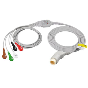
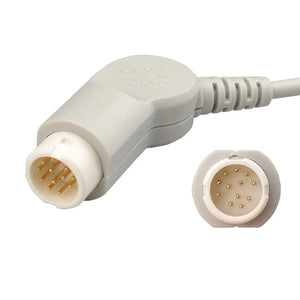
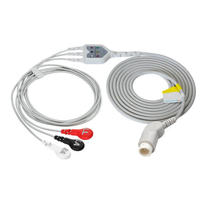
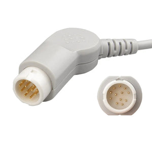
The 3-lead system uses three electrodes to capture leads I, II, and III, forming Einthoven’s Triangle—a horizontal projection of heart activity. It provides essential data on heart rate and rhythm. For reliable performance, consider the Philips M1669A trunk cable.

Key Advantages of 3-Lead ECG
- Easy to Use: Only three electrodes allow for quick setup, even for non-experts.
- Cost-Effective: Low cost makes it suitable for mass screenings or budget-constrained facilities.
- Real-Time Monitoring: Minimal signal delay ensures instant heart rate and ST-segment assessment.
Limitations
- Limited Scope: Misses vertical plane abnormalities, such as posterior wall infarction.
- Lower Accuracy: Misdiagnosis rates are 30–40% higher compared to 12-lead systems, according to NIH studies.
- Artifact Sensitivity: Motion artifacts or loose electrodes can distort readings.
Typical Applications of 3-Lead ECG
- Emergency Transport: Rapid heart monitoring during ambulance transport or disaster scenarios.
- Home Care: Long-term heart monitoring for chronic disease patients.
- Basic Screening: Early detection during general health checkups.
5-Lead ECG System: A Balanced Acute Care Solution
The 5-lead ECG system enhances diagnostic capabilities, making it a go-to choice for acute care monitoring.
Structure and Upgrades
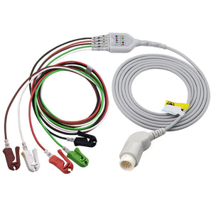
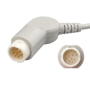
Building upon the 3-lead system, the 5-lead adds chest leads V1 and V6 to capture both horizontal and anterior/lateral heart activity. This broader diagnostic view increases sensitivity. The Philips M1668A trunk cable ensures reliable signal integrity for these setups.

Technical Advantages
- Broader Diagnostics: Detects right ventricular strain (V1) and lateral ischemia (V6), reducing missed diagnoses by 25% compared to 3-lead.
- Efficient Setup: Reuses some electrodes to limit total to five without sacrificing detail.
- Scalability: Fully compatible with 12-lead systems for seamless transitions from ER to inpatient care.
Typical Applications of 5-Lead ECG
- Emergency Departments: Immediate rhythm and ischemia assessment during chest pain episodes.
- ICUs: Continuous monitoring of unstable patients in critical care.
- Telemetry: Real-time inpatient monitoring.
“High-quality ECG cables and leadwires make the difference between a missed diagnosis and a saved life.” – Dr. Sarah Lin, Emergency Cardiologist
3-Lead vs. 5-Lead ECG: Quick Comparison
| Feature | 3-Lead ECG | 5-Lead ECG |
|---|---|---|
| Leads Displayed | I, II, III | I, II, III, V1, V6 |
| Electrodes | 3 | 5 |
| Best Use | Basic rhythm monitoring | Enhanced acute care diagnostics |
| Diagnostic Scope | Limited to horizontal plane | Includes anterior and lateral views |
Conclusion
Choosing between 3-lead and 5-lead ECG systems depends on clinical needs, but both rely on robust, reliable leadwires for accurate diagnostics. The 3-lead system, with products like the Philips M1669A, offers simplicity for basic rhythm monitoring, while the 5-lead system supported by the Philips M1668A delivers more comprehensive acute care diagnostics.
For technicians, distributors, and traders, investing in reliable ECG cables and leadwires ensures precision and patient safety across a wide range of clinical situations. High-quality cables remain the foundation for effective cardiac care.


