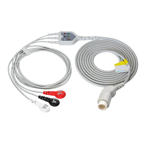
5 lead ECG placement can get messy when cables, colors, and chest landmarks collide. This streamlined guide keeps only what helps you move fast: where each electrode goes, how to choose the chest lead (V1 vs V5), the AHA↔IEC color map, essential skin prep/positioning, fast verification, and a short troubleshooting flow.
Quick-start map of the 5 leads
For continuous monitoring, place four “limb” electrodes on the torso to cut motion artifact and use one chest lead (V) for targeted ventricular insight.
- RA: Right upper chest, just below the clavicle (mid-clavicular), soft tissue.
- LA: Left upper chest, just below the clavicle (mid-clavicular), soft tissue.
- RL: Right lower torso, above the iliac crest.
- LL: Left lower torso, above the iliac crest.
- V (choose one): V1 = 4th ICS, right sternal border (rhythm); V5 = 5th ICS, anterior axillary line (ischemia/ST).
- Skin prep: clip hair, cleanse, dry; light abrasion per policy.
- Avoid bone, incisions, devices, irritated skin; press electrodes for 5–10 seconds.
- Confirm palette/labels (AHA vs IEC) before connecting leadwires.
- Provide strain relief: route cables downward and secure slack away from lines and dressings.
Safety: Use defibrillation-rated accessories when indicated; replace damaged cables/electrodes; follow infection-prevention policies.
Pick the chest lead: V1 vs V5
| Objective | Lead | Why |
|---|---|---|
| Arrhythmia / conduction | V1 | Best P-wave clarity; septal/right-sided view aids SVT vs VT and BBB patterns. |
| Ischemia / ST trending | V5 | Lateral wall sensitivity; reliable ST depression/elevation trends. |
Rule: One chest site at a time; label on the monitor and document the choice.
5-lead EKG Placement
byu/learningsponges inECG
Color coding: AHA vs IEC
Trust letters first, color second. This avoids cross-wiring when devices mix palettes.
| Lead | AHA | IEC | Label |
|---|---|---|---|
| RA | White | Red | RA / R |
| LA | Black | Yellow | LA / L |
| RL (ground) | Green | Black | RL / N |
| LL | Red | Green | LL / F |
| V (chest) | Brown | Brown | V / C |
Mnemonics that stick
- AHA: “White on right” (RA), “Clouds over grass” (white over green, right), “Smoke over fire” (black over red, left), “Chocolate near the heart” (V).
- IEC: “Red on right” (RA), “Sun over meadow” (yellow over green, left), RL black = neutral, V brown near the heart.
Tip: Use letters + color-blind-safe shapes on bedside cards; confirm palette in the monitor setup.
Prep & position (what actually improves signal)
- Skin: Clip hair; cleanse; dry fully; light abrasion per policy; press to warm gel.
- Position: Set posture first (supine or semi-Fowler’s 30–45°), then landmark ribs.
- Landmarks: Find the sternal angle → rib 2 → count to 4th/5th ICS (V1/V5).
Connect, verify, and monitor
- Seat connectors until they click; route cables down; secure slack away from lines.
- On the monitor: choose display lead (V1 rhythm / V5 ST), set gain/speed, minimize filters.
- Confirm trace: stable baseline, clear QRS, P waves when visible; capture a baseline strip and document chest site and palette (AHA/IEC).
Troubleshoot fast
-
Wandering baseline & motion
: Coach stillness; re-prep/dry skin; add slack; verify true 4th/5th ICS. -
Muscle noise & AC (mains) interference
: Warm patient, relax shoulders; separate ECG from power cords; ensure RL (ground) makes excellent contact. -
Lead-off alarms & loose/dry electrodes
: Replace electrode (don’t reuse), reseat leadwire, inspect pins/snaps; consider high-adhesion foam for sweat.
Product Compatibility & Buying Guide
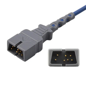
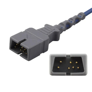
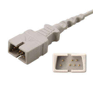
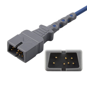
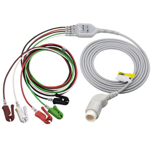
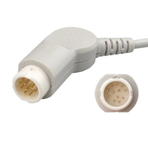
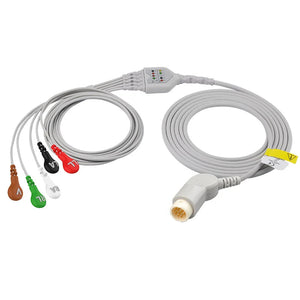
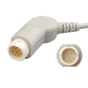
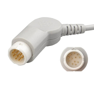
If prep is good but the signal is still noisy, cables are often the culprit. Replace leadwires for cracked jackets, corroded snaps/pins, or intermittent “wiggle” loss. Replace the trunk cable for broken strain reliefs, exposed conductors, or multi-lead dropouts that follow the trunk.
- Philips-compatible trunk cable M1668A
- Philips-compatible ECG leadwire M1625A
- Philips-compatible ECG leadwire M1623A
Pre-purchase fast check: match monitor port → trunk model → leadwire interface (snap/pin) → defib rating → cleaning compatibility → quick bedside trial.
Check compatibility or talk to our team about fleet matching.
Reference: American Heart Association (AHA). Additional reference: American College of Cardiology (ACC).
One-line takeaway: Prep dry skin, place limbs on soft tissue, pick V1 or V5 (and document), trust letters over colors, then verify a clean baseline before relying on alarms or trends.


