
📚 Series Article: This guide is part of our comprehensive 12 Lead ECG Placement: The Ultimate Guide. For standard placement technique, see our V1-V6 placement diagram.
Standard ECG placement guides assume an ideal patient: thin, cooperative, lying flat, with clear anatomical landmarks. Reality is different. Special patient ECG placement challenges arrive constantly—the morbidly obese patient whose ribs you can't feel, the post-cardiac surgery patient with sternal dressings, the COPD patient who can't lie flat, or the bilateral amputee with no limbs for standard electrode placement.
These situations require adaptation, not abandonment of technique. The principles remain constant: accurate electrode positioning matters for accurate diagnosis. But the methods of achieving accurate positioning must flex to meet each patient's unique anatomy and clinical circumstances.
This guide covers ECG placement for special situations you'll encounter regularly. For each scenario, you'll learn specific adaptations, documentation requirements, and the clinical reasoning behind the modifications.
⚡ Core Principle for All Special Situations
Document every adaptation. When you can't place electrodes in standard positions, note exactly what you did and why. This ensures interpreters understand any morphology changes and allows meaningful serial comparisons. A modified ECG with clear documentation is far more valuable than a "standard" ECG with unknown adaptations.
Special Situations Covered
- Obese Patients: Finding Landmarks
- Post-Surgical Patients: Working Around Dressings
- Respiratory Distress: Patients Who Can't Lie Flat
- Amputees: Alternative Limb Lead Placement
- Post-Mastectomy Patients
- Pediatric ECG Placement
- Chest Wall Deformities
- ICU and Ventilated Patients
- Patients with Implanted Devices
- Patients with Tremors or Movement Disorders
Obese Patients: Finding Landmarks When You Can't Feel Ribs
Obesity is the most common special ECG placement situation you'll encounter. Standard landmark palpation becomes difficult or impossible when subcutaneous tissue obscures bony anatomy.
The Challenge
- Angle of Louis: May be buried under tissue
- Intercostal spaces: Difficult to count
- Midclavicular line: Harder to identify with large breasts
- Breast tissue displacement: Affects V3-V6 positioning
- Skin folds: Poor electrode adhesion
Technique Adaptations
Finding the Angle of Louis
The standard technique starts at the sternal notch and moves down. For obese patients:
- Press firmly at the sternal notch—you should feel the top of the manubrium
- Slide your finger down the sternum with firm pressure
- The Angle of Louis is a horizontal ridge—it's often palpable even when ribs aren't
- If you can't feel it, use the nipple level approximation: in males, nipples are typically at the 4th-5th intercostal space (but verify if possible)
Comment
byu/SummChick from discussion
innursing
When ribs are impalpable:
- Find what you can feel—often the clavicle and lower ribs are palpable even when mid-chest isn't
- Count up from below: Find the lowest rib you can palpate, count up to estimate the 4th ICS
- Use the sternal border: Intercostal spaces are often easier to feel near the sternum than laterally
- Accept approximation: Document "estimated 4th ICS" rather than guessing without disclosure
Electrode Placement on Large Breasts/Abdomen
- V4-V6: Place at inframammary fold (under the breast) as described in our female patient ECG placement guide
- Lift and place: Gently lift breast/pannus tissue to place electrode on chest wall, then release
- Skin fold care: Separate skin folds, dry thoroughly, apply electrode to flat skin surface
Comment
byu/SignificantAccess905 from discussion
inbigboobproblems
Electrode Adhesion Issues
Obese patients often have moist skin in folds:
- Dry thoroughly with gauze before prep
- Use skin prep solution (benzoin tincture) for better adhesion if available
- Consider foam electrodes which conform better to uneven surfaces
- Tape edges if electrodes lift (ensure tape doesn't cover the gel)
📝 Documentation Example: "V1-V6 placed at estimated 4th and 5th ICS based on sternal palpation. Landmarks difficult to assess due to body habitus. V4-V6 placed at inframammary fold."
Post-Surgical Patients: Working Around Dressings and Drains
Post-cardiac surgery patients, thoracotomy patients, and those with chest tubes present unique ECG placement challenges.
Post-Sternotomy (CABG, Valve Surgery)
The sternal dressing often covers V1-V2 positions:
- Do NOT remove sterile dressings for ECG
- Place electrodes adjacent to dressing as close to correct position as possible
- Document the modification: "V1-V2 placed lateral to sternal dressing"
- If space allows: Some dressings have gaps that permit near-correct placement
Chest Tubes and Drains
Chest tubes often exit near V5-V6 positions:
- Never place electrode directly over tube site
- Identify the drain location
- Place electrode as close as safely possible without touching the drain
- Consider slight displacement (1-2 cm) rather than skipping the lead entirely
- Document: "V5 displaced 2cm superior due to chest tube"
Mastectomy or Breast Reconstruction
Fresh surgical sites require special care. For chronic post-mastectomy considerations, see the dedicated section below.
Pacemaker/ICD Implant Sites
Fresh generator sites (usually left infraclavicular) may affect upper limb lead positioning:
- Avoid placing electrodes over the fresh wound
- LA electrode may need slight displacement
- Document any modification
✓ Clinical Pearl: For serial ECG comparison in post-surgical patients, take a photo of your electrode positions (with patient consent) or draw a diagram. This allows the next clinician to replicate your exact placement.
Respiratory Distress: Patients Who Can't Lie Flat
Patients with heart failure, COPD exacerbation, or severe dyspnea often cannot tolerate supine positioning. This is a common special ECG placement situation that requires adaptation.
The Problem with Non-Supine ECGs
According to American Heart Association guidelines, patient position affects ECG morphology:
- QRS voltage: May decrease in upright position
- Axis: Shifts more vertical when sitting
- ST segments: Minor positional changes possible
These changes are usually minor but can affect serial comparison if position isn't documented.
Acceptable Alternate Positions
| Position | When to Use | Documentation |
|---|---|---|
| Semi-recumbent 30° | Mild dyspnea, can partially recline | "Semi-recumbent 30°" |
| Semi-recumbent 45° | Moderate dyspnea | "Semi-recumbent 45°" |
| Sitting upright | Severe dyspnea, orthopnea | "Seated upright—unable to recline" |
| High Fowler's | ICU patients, ventilated | "High Fowler's position" |
Technique for Seated Patients
- Landmark identification: Angle of Louis is still your starting point, but the chest wall shape changes when sitting
- Precordial leads: Place as normally as possible; gravity may shift breast tissue
- Limb leads: Can be placed on limbs in any position
- Arm support: Pillows under arms reduce tremor from muscle tension
- Document position precisely
Serial Comparison Considerations
When comparing ECGs taken at different positions:
- Expect some variation in voltage and axis
- Focus on significant changes (ST elevation, new Q waves) rather than subtle differences
- Try to match positions for serial comparison when possible
Amputees: Alternative Limb Lead Placement
Standard limb electrode placement requires intact limbs. Amputees need adapted ECG electrode placement strategies.
The Electrical Principle
The limb leads measure potential differences between points on the body surface. The key requirement is that electrodes be placed symmetrically and on the same type of tissue on each side.
Single Limb Amputation
Place the electrode on the residual limb (stump) as distally as possible:
- Below-knee amputation: Place LL electrode on remaining lower leg
- Above-knee amputation: Place LL electrode on thigh
- Below-elbow amputation: Place electrode on remaining forearm
- Above-elbow amputation: Place electrode on upper arm
Bilateral Lower Limb Amputation
When both legs are absent:
- Option 1: Place LL and RL electrodes on lower abdomen (below umbilicus), symmetrically
- Option 2: Place electrodes on the hips bilaterally
- Option 3: Use residual limbs if any length remains
Bilateral Upper Limb Amputation
- Place RA and LA electrodes on shoulders (anterior deltoid area)
- Ensure symmetric placement
Quadruple Amputee
Rare but encountered in trauma or severe peripheral vascular disease:
- RA/LA: Shoulders, symmetric
- RL/LL: Lower abdomen or hips, symmetric
📝 Documentation Example: "Bilateral BKA. LL electrode placed on left thigh, RL electrode placed on right thigh, symmetric positions. All subsequent ECGs should use same placement for comparison."
Critical Rule
Maintain symmetry. If you place LL on the left thigh, place RL on the right thigh—not one on thigh and one on abdomen. Asymmetric placement distorts the limb lead vectors.
Post-Mastectomy Patients
Mastectomy affects the left chest wall anatomy and requires adapted ECG placement techniques.
Unilateral Left Mastectomy
The chest wall anatomy changes:
- Landmarks more visible: Ribs and intercostal spaces may be easier to palpate
- V3-V6 placement: Use standard positions—no breast tissue to work around
- Scar tissue: Avoid placing electrodes directly on thick surgical scars (poor conductivity)
Bilateral Mastectomy
- Standard placement applies with no breast tissue modifications needed
- Avoid scar tissue when possible
- Document if scarring affects placement
Mastectomy with Reconstruction
Reconstructive surgery adds complexity:
- Implants: May change chest contour; use palpable landmarks
- TRAM/DIEP flaps: Different tissue thickness; may affect signal slightly
- Place electrodes on native chest wall tissue when possible
For general female patient considerations, see our female ECG placement guide.
Pediatric ECG Placement
Children are not small adults—pediatric ECG placement requires specific considerations.
Age-Related Differences
| Age Group | Electrode Size | Placement Considerations |
|---|---|---|
| Neonates | Neonatal/infant electrodes | Very small chest; electrodes may overlap |
| Infants (1-12 mo) | Pediatric electrodes | Small intercostal spaces |
| Children (1-8 yr) | Pediatric or small adult | Standard landmarks, scaled spacing |
| Adolescents | Adult electrodes | Adult technique applies |
Key Pediatric Differences
- Heart position: More horizontal in infants; shifts to adult orientation by age 7
- Intercostal spaces: Narrower; electrodes may be very close together
- Right ventricular dominance: Normal in infants; affects expected R-wave progression
Technique Tips
- Use appropriate electrode size—adult electrodes on infants cause overlap and artifact
- Landmarks are the same—4th ICS for V1-V2, just smaller
- Distraction helps: Toys, videos, or parental comfort reduce motion artifact
- Quick recording: Have everything ready before approaching child
- Warm electrodes: Cold electrodes on small children cause crying and artifact
Common Pediatric Artifacts
Children move. Expect and plan for:
- Motion artifact: Minimize with distraction, swaddling (infants)
- Crying artifact: Resembles severe muscle tremor; may need to wait or use filter
- Respiratory artifact: Faster respiratory rate causes more baseline variation
For artifact management techniques, see our ECG artifact troubleshooting guide.
Chest Wall Deformities
Structural chest abnormalities require adapted ECG electrode placement.
Pectus Excavatum (Funnel Chest)
The sternum is depressed inward:
- V1-V2: May be in the depression; place on the sloped chest wall
- Heart displacement: Heart may be shifted left; precordial leads see different orientation
- Standard positions apply but anatomy is shifted
- Document: "Pectus excavatum—standard electrode positions used"
Pectus Carinatum (Pigeon Chest)
The sternum protrudes outward:
- V1-V2: Standard sternal border positions, but chest wall angle differs
- Electrode adhesion: May be challenging on prominent sternum
- Document the deformity
Scoliosis/Kyphosis
Spinal curvature affects chest shape:
- Asymmetric chest: One side may be more prominent
- Use palpable landmarks as best you can
- Document: "Severe kyphoscoliosis—landmarks estimated"
Dextrocardia
Heart on the right side of the chest (congenital or acquired):
- Standard ECG shows: Inverted P, QRS, T in Lead I; poor R-wave progression
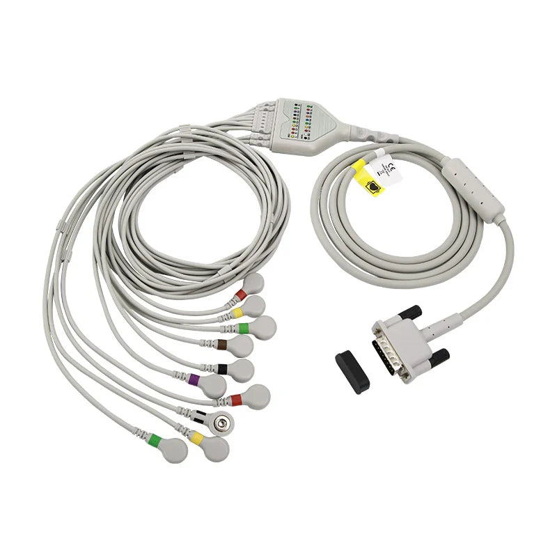
- Mirror-image placement: Place V1-V6 on the RIGHT side of chest—a complete 10-lead direct-connect cable ($96, Snap/IEC) gives you all six precordial leads to move across
- Label clearly: "Right-sided precordial leads—dextrocardia"
According to LITFL clinical resources, right-sided precordial placement in dextrocardia produces a "normal" appearing ECG that can be interpreted using standard criteria.
ICU and Ventilated Patients
Critical care patients present multiple ECG placement challenges simultaneously.
Common ICU Obstacles
- Central lines: Internal jugular, subclavian catheter dressings
- Arterial lines: Usually radial, occasionally femoral
- Chest tubes: One or multiple
- Surgical dressings: Sternal, thoracotomy
- Pacing wires: Epicardial wires post-cardiac surgery
- Monitoring electrodes: Already in place for continuous monitoring
- Ventilator tubing: May limit access
Practical Approach
- Survey the chest: Identify all obstacles before starting
- Plan electrode positions: Determine best achievable placement around obstacles
- Remove monitoring electrodes if needed: You can temporarily remove 3-lead or 5-lead monitoring electrodes to place 12-lead electrodes in their positions
- Work quickly: Minimize time off continuous monitoring
- Replace monitoring electrodes: After 12-lead is done
- Document everything
Ventilated Patients
- Respiratory artifact: Ventilator delivers consistent breaths; baseline may vary cyclically
- Position limitations: Often in semi-recumbent position (30-45°) for VAP prevention
- Sedation advantage: Sedated patients have less muscle tremor
Post-Cardiac Surgery Considerations
Multiple overlapping challenges:
- Sternal dressing covering V1-V2
- Chest tubes near V5-V6
- Pacing wires exiting near V3-V4
- Semi-recumbent position
Do your best, document thoroughly, and aim for consistency between serial ECGs.
Patients with Implanted Cardiac Devices
Pacemakers, ICDs, and loop recorders don't change ECG electrode placement technique, but they affect interpretation.
Pacemaker/ICD Placement
- Generator location: Usually left infraclavicular; avoid placing electrodes directly over the device
- Standard lead positions: Remain unchanged
- Paced rhythm: Will show pacing spikes and wide QRS (ventricular pacing)
What to Document
- Device present (pacemaker, ICD, CRT)
- Pacing activity observed (atrial, ventricular, dual-chamber)
- Any electrode displacement needed due to device
Loop Recorders
Implantable loop recorders (ILRs) are small and usually implanted in the left parasternal area:
- May be at V2-V3 location
- Place electrode adjacent to device if it's in the way
- Document the modification
Patients with Tremors or Movement Disorders
Parkinson's disease, essential tremor, and other movement disorders create significant ECG artifact challenges.
The Problem
Continuous involuntary movement causes:
- Persistent muscle tremor artifact
- Difficult to distinguish from atrial fibrillation
- May obscure fine ECG details
Management Strategies
- Optimize patient comfort: Warm room, comfortable position
- Support limbs: Pillows under arms and legs reduce transmitted tremor
- Time the recording: Some tremors are intermittent; wait for a quieter period
- Use filter settings: The "muscle" or "high-frequency" filter can reduce tremor artifact (use cautiously as it affects morphology)
- Accept the limitation: Document "ECG obtained with tremor artifact due to [condition]"
Distinguishing Tremor from Atrial Fibrillation
This is critical and covered in detail in our ECG artifact troubleshooting guide. Key points:
- Check RR intervals: Regular RR intervals suggest sinus rhythm with tremor artifact
- Look for P waves: May be visible between tremor oscillations
- Clinical correlation: Does the patient have known AF or a tremor condition?
Systematic Approach to Special ECG Placement Situations
Use this framework for any challenging ECG placement scenario:
Step 1: Assess the Situation (30 seconds)
- What obstacles are present? (dressings, drains, body habitus, position limitations)
- Which electrode positions are affected?
- Can the patient tolerate standard supine position?
Step 2: Plan Adaptations (30 seconds)
- Determine the closest achievable position for each electrode
- Decide on alternative positions if standard is impossible
- Prepare documentation language
Step 3: Execute with Documentation
- Place electrodes in the best achievable positions
- Document every adaptation immediately
- Note patient position
Step 4: Verify Quality
- Check for artifact
- Confirm all leads are recording
- Re-record if quality is inadequate
Special Situation Documentation Checklist
- ☐ Patient position documented (supine, semi-recumbent 30°, seated, etc.)
- ☐ Any electrode displacement noted with reason
- ☐ Obstacles described (dressings, drains, body habitus)
- ☐ Alternative placement positions specified if used
- ☐ Recommendation for serial comparison noted if applicable
🎯 Stock One Kit That's Ready for Any Patient
Adult + pediatric electrodes and a full 10-lead cable cover difficult adhesion, small chests, and right-sided / dextrocardia placement from a single cart
Frequently Asked Questions
Q: Should I skip an electrode if I can't place it in the correct position?
A: No. Always place every electrode somewhere reasonable. A slightly displaced electrode provides more information than a missing one. Document the displacement. The only exception is if placing the electrode would cause harm (over a sterile surgical site, for example).
Q: How important is documentation of modifications?
A: Critical. An ECG interpreter who doesn't know V2 was displaced may misinterpret morphology changes as pathological. Documentation protects the patient and allows meaningful comparison. Write the modifications directly on the ECG and in the medical record.
Q: Can I use a 6-lead ECG instead of 12-lead if the patient is too difficult?
A: A 6 lead ECG placement (limb leads only) can be done as a fallback, but it provides much less information than 12-lead. It misses the precordial leads entirely, which assess the anterior, septal, and lateral walls. Always attempt 12-lead first. Use 6-lead only when precordial leads are truly impossible.
Q: What if the patient can't stay still due to pain?
A: Address the pain first if possible. A brief delay for pain medication often produces a better quality ECG. If you must record immediately, do your best and document: "ECG obtained with significant motion artifact due to uncontrolled pain."
Mastering Special Patient ECG Placement
Special patient ECG placement challenges are not exceptions—they're daily reality. Obesity, post-surgical states, respiratory distress, and other special situations account for a significant proportion of the ECGs you'll perform.
The key principles apply across all special ECG placement situations:
- Adapt the technique, not the standards: The goal remains accurate electrode positioning; the method changes
- Document everything: Every modification, displacement, and limitation
- Maintain symmetry: Especially for limb leads in amputees or alternative placements
- Note patient position: Essential for serial comparison
- Some ECG is better than no ECG: A modified recording with documentation beats waiting for "perfect" conditions
Master these ECG placement adaptations, and you'll be prepared for any patient who walks (or is wheeled) through your door.
📚 Complete Your ECG Placement Skills
Return to the main guide: 12 Lead ECG Placement: The Ultimate Guide
Related guides for challenging situations:
Clinical Education Team
Our clinical team works in environments where "textbook" patients are rare. This guide reflects real-world experience with thousands of ECGs performed on patients with challenging anatomy, surgical modifications, and complex clinical situations.


