
12 lead ECG placement for female patients is a topic that most training programs barely mention—yet it's one of the most common sources of diagnostic error I encounter. The reality is that breast tissue significantly affects electrode positioning and signal quality, and failing to address this can lead to misdiagnosis.
I learned this lesson from a 45-year-old woman whose ECG showed flattened T-waves and what appeared to be ischemic changes. After repositioning V4-V6 from on top of her breast tissue to underneath it, her ECG was completely normal. The "ischemia" was simply signal attenuation from breast tissue blocking the electrical signal.
This guide covers everything you need to know about ECG lead placement female patients: why breast tissue matters, exactly where to position electrodes, how to communicate professionally, and the techniques that ensure accurate, reproducible results.
⚡ Key Takeaways
- Breast tissue can attenuate ECG signals by 30-40%, creating false abnormalities
- For patients with larger breasts, V4-V6 should go under the breast at the inframammary fold
- For smaller breasts, electrodes can be placed on top of the breast without significant signal loss
- Professional communication and privacy measures are essential
- Always document your approach for serial ECG comparison
Quick Reference: Over the Breast or Under?
When in doubt, under-breast placement is the safer choice for signal quality.
In This Guide
Why Female ECG Placement Requires Special Attention
Standard ECG training typically shows electrode placement on a flat male chest. But approximately half of your patients will be female, and many will have breast tissue that directly affects where V3, V4, V5, and V6 should be positioned.
The challenge is straightforward: the precordial leads need to be placed at specific anatomical landmarks (the 4th and 5th intercostal spaces), but breast tissue may cover these exact locations. You have two options—place the electrodes on the breast, or place them under the breast—and each choice has implications.
What the Research Shows
According to guidelines from the American Heart Association and other major cardiology organizations, there is no universal consensus on whether electrodes should go over or under breast tissue. However, the clinical evidence strongly suggests that significant breast tissue causes signal attenuation when electrodes are placed on top.
The bottom line: you need to assess each patient individually and make an informed decision based on breast size and tissue density.
How Breast Tissue Affects ECG Signals
Understanding the physics helps explain why placement matters so much for woman 12 lead ECG placement.
The Signal Attenuation Problem
Breast tissue acts as an electrical insulator. When electrodes are placed on top of dense or thick breast tissue, the electrical signals from the heart must pass through this additional layer before reaching the electrode. This causes:
- Reduced R-wave amplitude: The QRS complexes appear smaller than they actually are
- Altered T-wave morphology: T-waves may appear flattened or inverted
- Apparent low voltage: The entire ECG may look "dampened"
- False positive findings: Changes that mimic ischemia or other pathology
Research published in clinical cardiology journals indicates that breast tissue can reduce signal amplitude by 30-40% in some cases. That's enough to completely change the interpretation of an ECG. It's worth noting that cable quality also plays a role — degraded cable shielding and impedance can compound signal attenuation from tissue, making it even harder to distinguish true pathology from artifact. This is why it helps to understand how connector fit, pinout, and signal stability are bench-verified — an unstable connector mate or noisy contact stacks on top of tissue attenuation and makes a clean tracing even harder to obtain.
A Case That Illustrates the Problem
A 45-year-old woman came to our department with atypical chest discomfort. Her initial ECG showed:
- Flattened T-waves in V4-V6
- Low voltage in lateral leads
- Pattern suggesting possible lateral ischemia
The cardiology team was considering admission for further workup. But something didn't fit—her symptoms were atypical, and she had no cardiac risk factors.
I noticed that V4-V6 had been placed directly on top of her breast tissue. I repositioned them at the inframammary fold (where the breast meets the chest wall) and repeated the ECG.
The result: completely normal T-waves, normal voltage, no ischemic changes.
The "ischemia" was artifact from signal attenuation. She went home that day instead of being admitted for a cardiac workup she didn't need.
The Placement Decision: Over the Breast or Under?
This is the key question for ECG lead placement female patients. The answer depends on breast size and tissue characteristics.
When to Place Electrodes ON TOP of the Breast
Appropriate for: Patients with small to medium breasts where tissue thickness is minimal
If the breast tissue is thin enough that you can easily palpate the underlying ribs and intercostal spaces through it, placing electrodes on top is acceptable. The tissue layer isn't thick enough to cause significant signal attenuation.
Advantages:
- Maintains standard anatomical positioning
- May be more comfortable for the patient
- Easier and faster to apply
When to Place Electrodes UNDER the Breast
Appropriate for: Patients with larger breasts or dense breast tissue
According to LITFL's clinical guidelines and other expert resources, when breast tissue is substantial, electrodes V4-V6 should be placed at the inframammary fold—the crease where the breast meets the chest wall.
Advantages:
- Direct contact with chest wall
- No signal attenuation from breast tissue
- More accurate ECG representation
Note: This means V4-V6 may end up slightly lower than the strict "5th intercostal space" definition, but the improved signal quality outweighs the minor positional variation.
Quick Decision Guide
Can you easily palpate the 5th intercostal space through the breast tissue?
- YES → Place electrodes in standard position (on breast is acceptable)
- NO → Place V4-V6 at the inframammary fold (under the breast)
When in doubt: Under the breast is the safer choice for signal quality.
Step-by-Step ECG Lead Placement for Female Patients
Here's my systematic approach for 12 lead ECG placement female patients, refined over years of clinical practice.
Assessment
Before placing any electrodes, assess:
- Breast size and tissue density
- Whether you can palpate landmarks through breast tissue
- Any surgical scars, implants, or other considerations
- Patient's comfort level and any concerns
V1 and V2 (Usually Unaffected)
V1 and V2 are placed at the sternal borders in the 4th intercostal space. For most women, breast tissue doesn't extend this far medially, so standard placement applies. For the detailed V1-V2 technique, see our 12 lead ECG placement diagram guide.
V4 Position Decision
V4 is where the decision point occurs. Assess whether V4's standard position (5th intercostal space, midclavicular line) falls on breast tissue.
- If minimal breast tissue at V4 location: Place in standard position
- If significant breast tissue at V4 location: Place at the inframammary fold, directly below the standard V4 position
V3 Positioning
V3 goes halfway between V2 and V4. If you've moved V4 to the inframammary fold, V3 will also shift accordingly to maintain the midpoint relationship.
V5 and V6 Positioning
V5 and V6 must be at the same horizontal level as V4. If V4 is at the inframammary fold, V5 and V6 should also follow that horizontal line.
For patients where you're placing under the breast:
- Gently lift the breast or ask the patient to hold it
- Identify the inframammary fold
- Place V4 at the midclavicular line along the fold
- Place V5 and V6 laterally, keeping them horizontal with V4
- Release the breast—electrodes should remain in place
Final Check
Verify:
- V1-V2 are level with each other
- V4-V5-V6 form a horizontal line
- All electrodes have good skin contact
- Patient is comfortable
If the ECG still shows unexpected low voltage or artifact after confirming proper placement, the issue may be equipment-related. Worn lead wire snap connectors or a damaged trunk cable can mimic the signal attenuation caused by breast tissue. Replacing electrodes first is the right clinical step, but if problems persist, ensure your ECG cable connector type matches your monitor and that cables pass a visual cable inspection and testing check.

Professional Communication Techniques
How you communicate about this sensitive topic significantly affects patient comfort, cooperation, and the quality of the ECG you obtain.
Before Starting: Set Expectations
My approach:
"I'm going to place some electrodes on your chest to record your heart's electrical activity. I'll need to place some of them below your breast to get the most accurate reading. Is that okay with you?"
This script accomplishes several things:
- Explains what you're doing
- Gives advance notice about breast area involvement
- Asks for consent
- Uses professional, clinical language
During the Procedure
Narrate your actions briefly:
- "I'm going to place this electrode here..."
- "I need to position this one under your breast..."
- "Almost done—just two more..."
This keeps the patient informed and reduces anxiety about what's happening.
If the Patient Has Questions or Concerns
Be prepared to explain:
- Why placement under the breast matters: "Breast tissue can interfere with the signal, so placing the electrode underneath gives us a clearer picture of your heart."
- That this is standard practice: "This is the recommended technique for accurate results."
- That you'll maintain their dignity: "I'll keep you covered as much as possible and work quickly."
Privacy and Dignity Considerations
Maintaining patient dignity is non-negotiable. Here's how I approach it:
Environmental Setup
- Close the door and curtain before any exposure
- Check for unexpected interruptions—put a sign on the door if needed
- Ensure adequate lighting so you can work efficiently without prolonged exposure
Draping Technique
- Provide a gown or sheet for the patient
- Expose only the areas you're actively working on
- Use a towel or small drape to cover areas where electrodes are already placed
- Work systematically to minimize total exposure time
Chaperone Considerations
Depending on your facility's policy and the patient's preference:
- Offer a same-gender clinician if available and preferred
- Offer a chaperone to be present
- Document the patient's preference and your actions
✓ Best Practice: Ask the patient if they would like a chaperone or a female staff member present. This demonstrates respect for their comfort and protects both patient and clinician.
Documentation Requirements for Female Patient ECGs
Proper documentation is essential for ECG comparison and clinical accuracy.
What to Document
When you modify standard placement for breast tissue, note it on the ECG:
- "V4-V6 placed at inframammary fold"
- "Electrodes placed under breast tissue per protocol"
- "V3-V6 positioned below breast—standard landmarks obscured"
Why Documentation Matters
- Serial comparison: If subsequent ECGs are done with different placement, changes might be positional rather than pathological
- Interpretation accuracy: The reading physician needs to know about any modifications
- Reproducibility: Other clinicians can replicate your approach for consistent monitoring
For more on quality documentation practices, see our ECG quality control system guide.
Special Situations
Breast Implants
Breast implants present similar challenges to natural breast tissue:
- Silicone and saline both attenuate electrical signals
- Place electrodes at the inframammary fold, below the implant
- Document: "V4-V6 placed below breast implant"
Post-Mastectomy Patients
For patients who have had a mastectomy:
- Place electrodes in standard anatomical positions on the affected side
- Be sensitive to surgical scars and patient comfort
- Document the surgical history and any placement modifications
Asymmetric Breasts
When breast size differs significantly between sides:
- Assess each side independently
- You may use different approaches for right-sided vs. left-sided leads
- Document your approach for each side
Patients Who Decline Modified Placement
Occasionally, a patient may be uncomfortable with electrode placement under the breast. In this case:
- Respect their wishes
- Place electrodes in standard position on top of breast tissue
- Document: "Patient preference—electrodes placed over breast tissue"
- Alert the interpreting physician to potential signal attenuation
Pregnancy Considerations
During pregnancy, breast tissue often increases significantly. The same principles apply—assess tissue thickness and use inframammary placement when appropriate.
For other challenging patient populations, see our guide on special ECG placement situations.
Frequently Asked Questions
Should electrodes always go under the breast for female patients?
No. The decision depends on breast size and tissue density. For patients with smaller breasts where you can easily palpate the underlying ribs, standard placement on top is acceptable. For larger breasts or dense tissue, placement at the inframammary fold provides better signal quality.
Does breast size affect ECG interpretation?
It can, if electrodes are placed on top of significant breast tissue. The tissue attenuates signals, potentially causing false-positive findings like low voltage or T-wave changes. Proper placement technique eliminates this issue.
What if the patient is uncomfortable with under-breast placement?
Respect their preference and document it. Place electrodes in standard position, note "patient preference—electrodes placed over breast tissue," and alert the interpreting physician to potential signal attenuation. Patient autonomy takes priority.
Do I need a female clinician to perform ECGs on female patients?
This depends on your facility's policy and patient preference. Best practice is to offer a same-gender clinician or chaperone if available and desired. Always ask the patient what they prefer and document accordingly.
Ensuring Accurate ECGs for Every Female Patient
Proper 12 lead ECG placement for female patients isn't complicated once you understand the principles. The key is recognizing that breast tissue affects signal quality, making an informed decision about electrode positioning, communicating professionally, and documenting your approach.
The techniques in this guide for ECG lead placement female patients will help you avoid the diagnostic errors that come from signal attenuation—like the "ischemia" that disappears when you simply move the electrodes to the right location.
Remember: every patient deserves an accurate ECG. Taking the extra few seconds to assess and properly position electrodes can prevent unnecessary cardiac workups, reduce patient anxiety, and ensure that real pathology doesn't get missed or invented.
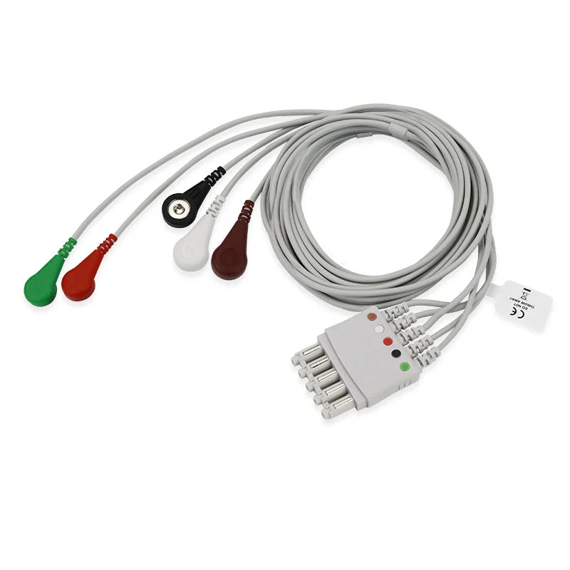
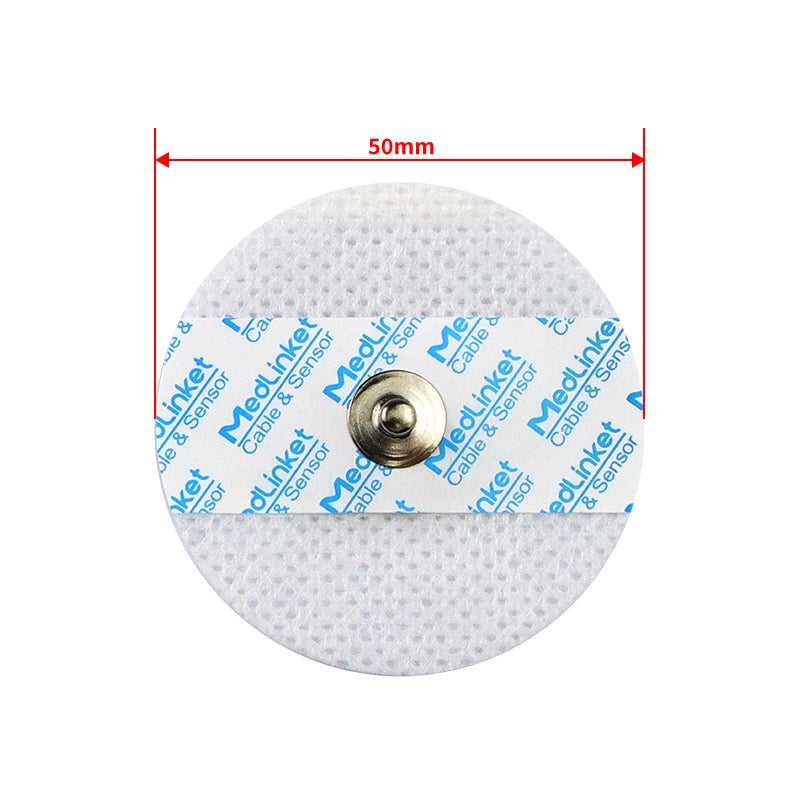
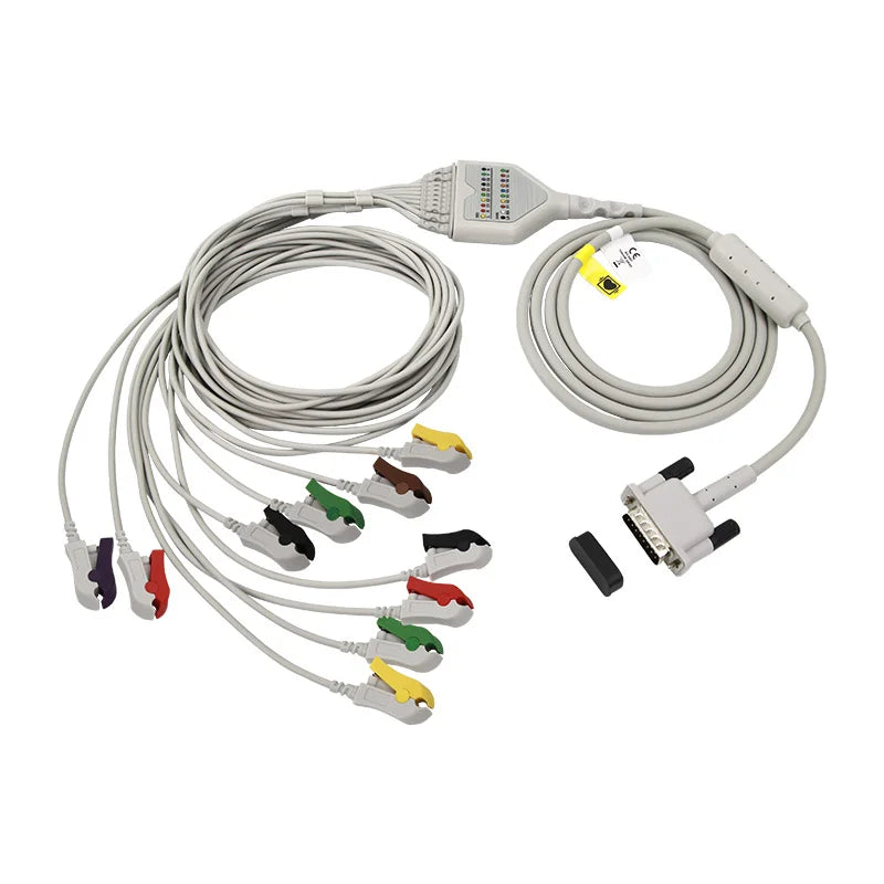
Ensure Signal Quality with the Right ECG Accessories
Accurate ECG interpretation depends on both proper electrode placement AND reliable accessories. MedLinket manufactures a complete line of ECG cables, leadwires, and electrodes compatible with Philips, GE, Mindray, Nihon Kohden, Schiller, and Mortara monitors.
Serving 2,000+ hospitals across 120+ countries · ISO 13485 · FDA & CE certified
Free monitor compatibility verification: shopify@medlinket.com
Clinical Education Team
Our clinical education team includes experienced emergency nurses, cardiac technicians, and cardiology specialists — with women clinicians involved in reviewing all guidance that touches on female patient care. This article reflects AHA guidelines, LITFL clinical best practices, and bedside techniques validated across U.S. emergency departments and cardiac units to ensure both diagnostic accuracy and patient dignity.


