
Looking for a reliable 12 lead ECG placement mnemonic to remember electrode positions? You're not alone. Whether you're a nursing student preparing for clinicals, a paramedic reviewing for recertification, or a seasoned clinician who just needs a quick refresher, memory tricks like "White on Right", "Smoke Over Fire", and "Snow Over Grass" make ECG placement faster and more accurate.
This guide compiles the most effective ECG placement mnemonics used in U.S. hospitals (AHA color system), answers frequently asked questions about electrode positioning, and provides quick reference charts you can use at the bedside. Bookmark this page—you'll want to come back to it.
🔍 Quick Jump
In This Guide
Limb Lead Placement Mnemonics
The four limb electrodes form the foundation of the 12-lead ECG. Here are the most popular memory tricks for getting them right every time.
AHA System: "White on Right, Smoke Over Fire"
This is the most widely used ECG placement mnemonic in U.S. hospitals:
🇺🇸 AHA Color Mnemonic (United States)
- "White on Right" → White electrode on Right Arm
- "Smoke Over Fire" → Black (smoke) above Red (fire) on left side
- "Snow Over Grass" → White (snow) above Green (grass) on right side
- Green = Ground → Green on Right Leg (ground/reference electrode)
The Classic: "Ride Your Green Bike"
Another widely taught 12 lead ECG placement mnemonic:
🚲 "Ride Your Green Bike"
- Ride = Red → Right Arm (RA)
- Your = Yellow → Left Arm (LA)
- Green = Green → Right Leg (RL)
- Bike = Black → Left Leg (LL)
IEC System: "Red Right, Yellow Left" + Traffic Light
For the International Electrotechnical Commission color coding (used outside the US):
🌍 IEC Color Mnemonic (International)
- "Red Right" → Red on Right Arm
- "Yellow Left" → Yellow on Left Arm
- "Traffic Light on Legs" → Green (go) on Left Leg, Black on Right Leg
For detailed limb lead placement technique, see our 5 lead ECG placement guide.
Precordial Lead (V1-V6) Placement Mnemonics
The precordial leads require precise anatomical positioning. These 12 lead ECG placement mnemonics help you remember the locations.
The Landmark Mnemonic: "2-4-5 Rule"
📍 The 2-4-5 Rule
- V1 & V2: Start at 2nd rib (Angle of Louis), drop to 4th intercostal space
- V4: 5th intercostal space, midclavicular line
- V3: Halfway between V2 and V4
- V5 & V6: Same horizontal level as V4
"V1 V2 at the Sternum, V4 at the Nipple"
A simple anatomical reminder:
- V1: Right sternal border, 4th ICS
- V2: Left sternal border, 4th ICS
- V4: Midclavicular line, 5th ICS (approximately nipple level in males)
⚠️ Caution: The "nipple level" reference works for males but NOT for females. For female patients, always use anatomical landmarks (5th ICS, midclavicular line). See our female patient ECG placement guide.
V5-V6 Horizontal Mnemonic: "V4-5-6 Stay Level"
The most common precordial placement error is letting V5 and V6 drift upward. Remember:
✓ V4-5-6 Rule
V4, V5, and V6 must be on the same horizontal line. If V4 is in the 5th ICS, then V5 and V6 are also in the 5th ICS—even as you move laterally around the chest.
For a complete visual guide, see our 12 lead ECG placement diagram.
ECG Electrode Color Codes: AHA vs IEC
Different countries use different color systems. According to the American Heart Association, the AHA system is standard in the United States, while the IEC system is used internationally. When ordering replacement ECG cables, matching the correct color standard to your region is essential — our ECG cable connector types technical guide covers both AHA and IEC cable variants across all major monitor brands.
Complete Color Code Reference Table
| Electrode | Position | AHA (USA) | IEC (International) |
|---|---|---|---|
| RA | Right Arm | White | Red |
| LA | Left Arm | Black | Yellow |
| RL | Right Leg | Green | Black |
| LL | Left Leg | Red | Green |
| V1-V6 | Precordial | Brown (both systems) — or individually color-coded | |
Quick Memory Trick for System Differences
- AHA: "White and Green on the Right" (White-RA, Green-RL)
- IEC: "Red on Right arm, Green on Left leg" (traffic light colors on opposite corners)
Best Order for ECG Electrode Placement
While there's no single "correct" order, this sequence minimizes errors and improves efficiency:
Recommended Placement Sequence
- Prepare skin at all 10 electrode sites first
- Limb leads — RA → LA → RL → LL (clockwise from patient's right arm)
- V1 — Right sternal border, 4th ICS (find Angle of Louis first)
- V2 — Left sternal border, 4th ICS (directly across from V1)
- V4 — 5th ICS, midclavicular line (skip V3 for now)
- V3 — Halfway between V2 and V4 (now you can measure exactly)
- V5 — Same level as V4, anterior axillary line
- V6 — Same level as V4, midaxillary line
- Verify — Check all connections and electrode adhesion
- Record
Why Skip V3 Initially?
V3 is defined as the midpoint between V2 and V4. If you place V4 first, you can precisely measure where V3 should go. "Eyeballing" V3 without V4 as a reference is a common ECG placement mistake.
Basic ECG Placement FAQs
What are the 10 ECG electrode placement positions?
- 4 Limb electrodes: Right Arm (RA), Left Arm (LA), Right Leg (RL), Left Leg (LL)
- 6 Precordial electrodes: V1, V2, V3, V4, V5, V6 on the chest
Where exactly is the 4th intercostal space?
Start at the sternal notch (top of the sternum). Slide your finger down until you feel a horizontal ridge—this is the Angle of Louis, which marks the 2nd rib. The space below the 2nd rib is the 2nd intercostal space. Count down two more spaces to reach the 4th intercostal space, where V1 and V2 are placed.
Why does a "12-lead" ECG only use 10 electrodes?
The term "12-lead" refers to 12 different electrical views of the heart, not 12 physical electrodes. The 4 limb electrodes generate 6 leads (I, II, III, aVR, aVL, aVF) through various combinations. The 6 chest electrodes each generate one lead (V1-V6). Total: 6 + 6 = 12 leads from 10 electrodes.
What is the difference between 3-lead, 5-lead, and 12-lead ECG?
- 3-lead: Uses 3 electrodes; shows only Lead I, II, and III; used for basic rhythm monitoring
- 5-lead: Uses 5 electrodes; shows limb leads plus one chest lead; used for continuous monitoring
- 12-lead: Uses 10 electrodes; shows all 12 leads; used for complete cardiac assessment
Does it matter if the patient is lying down or sitting up?
Yes. The standard position is supine (lying flat). Sitting or semi-recumbent positions can slightly alter QRS voltage and axis. If the patient cannot lie flat, document the position on the ECG. For details, see our special situations guide.
Troubleshooting FAQs
What causes baseline wander on ECG?
Baseline wander is usually caused by poor electrode contact. Common causes include: inadequate skin preparation, dried electrode gel, patient movement (especially breathing), loose electrode adhesion, or sweaty skin. Fix by re-prepping skin and using fresh electrodes. See our complete ECG artifact troubleshooting guide.
How do I tell if leads are reversed?
The classic sign of LA-RA (arm lead) reversal is an inverted P wave and QRS in Lead I combined with an upright P and QRS in aVR. This pattern should immediately trigger a check of lead connections. For other reversal patterns, see our 10 common ECG placement mistakes.
What does "artifact" mean on an ECG?
Artifact is any electrical signal on the ECG that doesn't come from the heart. Sources include muscle movement, electrical interference (60 Hz), poor electrode contact, and patient motion. Artifacts can mimic arrhythmias and cause misdiagnosis, so it's important to identify and eliminate them.
Why is my ECG showing flat lines in some leads?
Flat lines usually indicate a disconnected electrode or broken cable. Check that: (1) the electrode is firmly attached to the skin, (2) the cable clip is fully connected to the electrode, and (3) the cable isn't damaged. If one lead is flat, the problem is that specific electrode. If all leads are flat, check the machine connection. Persistent flat-line issues after replacing electrodes may point to a faulty lead wire or trunk cable — different monitors use different ECG cable connector types, so ensure the replacement matches your specific model. If you're running a Philips IntelliVue MP-series monitor, you can confirm the right leadwire and cable on our Philips IntelliVue MP-series compatible accessories page.
What causes 60 Hz interference on ECG?
60 Hz (or 50 Hz outside the US) interference comes from nearby electrical sources. Common culprits: cell phones near the patient, IV pumps touching the bed, fluorescent lights, and poor grounding. Move electronic devices away, ensure good RL (ground) electrode contact, and unplug unnecessary equipment. If interference persists after eliminating environmental sources, the ECG cable's internal shielding may be compromised — proper cable shielding and impedance is what prevents external electromagnetic interference from contaminating the signal.
Special Situations FAQs
Where do I place electrodes on female patients?
Place V3-V6 electrodes under the left breast (at the inframammary fold), not on top of breast tissue. The electrodes should contact the chest wall directly. Placing electrodes on breast tissue causes signal attenuation and inaccurate readings. See our complete female patient ECG placement guide.
How do I place ECG electrodes on obese patients?
Use firm pressure to palpate landmarks (the Angle of Louis is often still palpable). If you can't feel ribs, count up from lower ribs that you can palpate, or use the sternal border where intercostal spaces are easier to feel. Document "estimated positions" if landmarks are unclear.
What is V4R and when should I use it?
V4R is a right-sided chest lead, placed on the right side of the chest in the mirror position of V4. It's used to detect right ventricular infarction, especially in patients with inferior STEMI. According to the European Society of Cardiology, V4R should be obtained in all patients with inferior STEMI. See our right-sided ECG lead placement guide.
When should I use posterior leads (V7-V9)?
Posterior leads help diagnose posterior myocardial infarction. Use them when you see ST depression in V1-V3 (which may be a "mirror image" of posterior ST elevation) or when clinical suspicion is high but the standard 12-lead is non-diagnostic. See our posterior ECG lead placement guide.
What is a 15-lead ECG?
A 15-lead ECG adds three additional leads to the standard 12: V4R (right ventricular view) and V8-V9 (posterior views). It's used when there's concern for right ventricular or posterior involvement, particularly in patients with inferior STEMI. See our 15-lead ECG placement guide.
Can I place ECG electrodes over a pacemaker?
Avoid placing electrodes directly over the pacemaker generator (usually in the left infraclavicular area). Place electrodes adjacent to the device. The pacemaker won't interfere with the ECG, but you'll see pacing spikes on the tracing.
Quick Reference Card: 12 Lead ECG Placement Mnemonic Summary
Print or screenshot this quick reference for bedside use:
📋 ECG QUICK REFERENCE CARD
LIMB LEADS (AHA)
- RA: White — Right Arm
- LA: Black — Left Arm
- RL: Green — Right Leg
- LL: Red — Left Leg
Memory: "White on Right, Smoke Over Fire"
PRECORDIAL LEADS
- V1: 4th ICS, R sternal border
- V2: 4th ICS, L sternal border
- V3: Between V2 and V4
- V4: 5th ICS, midclavicular
- V5: V4 level, ant. axillary
- V6: V4 level, mid-axillary
KEY RULES
- ✓ Start at Angle of Louis (2nd rib) to find 4th ICS
- ✓ V4-V5-V6 must be on the same horizontal line
- ✓ Place V3 AFTER V4 (so you can measure the midpoint)
- ✓ Females: V3-V6 under the breast, not on it
💡 Press Ctrl+P (or Cmd+P) to print this page · Screenshot for mobile bedside reference
Top 5 ECG Placement Errors to Avoid
Based on clinical audits, these are the most frequent mistakes. For detailed analysis, see our 10 common ECG placement mistakes guide.
| # | Error | Quick Fix |
|---|---|---|
| 1 | V1-V2 placed too high (2nd or 3rd ICS) | Always start at Angle of Louis |
| 2 | V5-V6 not level with V4 | Use ruler or string to maintain level |
| 3 | Electrodes on breast tissue | Place under the breast |
| 4 | LA-RA lead reversal | Check Lead I and aVR before accepting |
| 5 | Poor skin preparation | Clean, abrade until pink, dry |
Master ECG Placement with Mnemonics
A reliable 12 lead ECG placement mnemonic transforms a complex skill into automatic muscle memory. Whether you prefer "White on Right", "Smoke Over Fire", or the "2-4-5 Rule", the key is choosing one system and practicing it consistently until it becomes second nature.
These ECG placement mnemonics and FAQs cover the questions we hear most often from clinicians at all experience levels. Bookmark this page for quick reference, and remember:
- Anatomy trumps memory tricks — When in doubt, verify landmarks
- Practice builds speed — Use our ECG quality control system for structured training
- Document variations — Any deviation from standard placement should be noted
- Check before accepting — Lead I and aVR catch most reversal errors
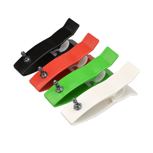
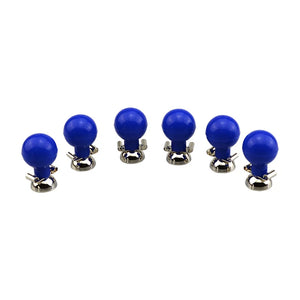
With the right 12 lead ECG placement mnemonic and consistent technique, you'll perform accurate ECGs quickly and confidently—every time. If your department is also restocking the consumables behind these placements, our AHA-coded limb clamp electrodes ($40, 20% off) and 21mm precordial suction electrodes ($40, 20% off) cover both the limb and chest positions described above.
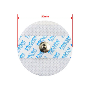
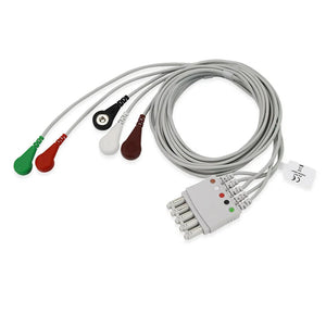
Complete ECG Setup? Check Your Accessories
Correct mnemonics help you place electrodes in the right positions — but signal quality depends equally on your ECG cables and electrodes. MedLinket manufactures a complete line of ECG accessories compatible with Philips, GE, Mindray, Nihon Kohden, Schiller, and Mortara monitors used in U.S. hospitals.
Serving 2,000+ hospitals across 120+ countries · ISO 13485 · FDA & CE certified
Free monitor compatibility verification: shopify@medlinket.com
Clinical Education Team
Our team includes emergency nurses, paramedics, and cardiac technicians with combined decades of experience training clinical staff on ECG technique. These mnemonics reflect the memory tricks that actually stick — compiled from classroom teaching, bedside practice, and feedback from nursing students, EMT programs, and hospital CE sessions across the United States.


