
📚 Series Article: This guide is part of our comprehensive 12 Lead ECG Placement: The Ultimate Guide. For standard chest lead positioning, see our V1-V6 placement diagram first.
Right sided ECG lead placement is one of the most critical—and most commonly skipped—steps in acute cardiac care. If you see an inferior STEMI and don't check V4R, you're practicing incomplete medicine. I'll be direct about that.
June 2023. A 68-year-old man arrived with classic inferior STEMI—ST elevations in leads II, III, and aVF. His blood pressure was borderline at 90/60. The standard treatment for inferior MI? Nitroglycerin for pain relief.
But something felt wrong. I ran a V4R. It showed 5mm of ST elevation—massive right ventricular infarction.
If we had given nitroglycerin based on the standard 12-lead alone, his preload-dependent right ventricle would have crashed. The nitro would have dropped his blood pressure to dangerous levels. Instead, we modified treatment immediately: IV fluids, no nitrates, and a different cath lab approach.
He survived. But this case is why I now perform right sided ECG lead placement on every inferior STEMI. No exceptions.
🚨 Critical Clinical Rule
Every inferior STEMI requires V4R. Right ventricular involvement occurs in 30-50% of inferior MIs and completely changes management. Missing it can be fatal.
In This Guide
When Right Sided ECG Lead Placement Is Mandatory
Right sided leads aren't routine—they're indicated in specific clinical scenarios. Here's when you must perform them:
Absolute Indications for V4R
✓ Always check V4R when you see:
- Inferior STEMI (ST elevation in leads II, III, aVF) — Every single time
- Inferior MI with hypotension — Especially if BP drops with nitrates
- ECG inferior leads showing ST elevation with III > II pattern
- Suspected right ventricular involvement from any cause
The Statistics That Should Convince You
According to American Heart Association data and clinical research:
- 30-50% of inferior STEMIs involve the right ventricle
- Right ventricular MI increases in-hospital mortality by 25-30%
- V4R alone has 88% sensitivity for detecting RV infarction
- Standard 12-lead ECG misses isolated RV involvement entirely
These aren't obscure statistics—this is the difference between appropriate treatment and potential harm.
Recognizing ECG Inferior Leads Pattern
Before discussing right sided placement, ensure you can identify the inferior STEMI that triggers this protocol:
- Lead II: ST elevation
- Lead III: ST elevation (often greater than Lead II in RV involvement)
- Lead aVF: ST elevation
- Reciprocal changes: ST depression in leads I and aVL
When you see this pattern, V4R becomes mandatory—not optional.
Understanding Right Ventricular Infarction
To understand why right sided ECG lead placement matters, you need to understand what happens when the right ventricle infarcts.
The Anatomy
The right coronary artery (RCA) supplies:
- The inferior wall of the left ventricle
- The right ventricle
- The SA and AV nodes (in most people)
When the RCA occludes proximally, it can damage both the inferior LV wall AND the right ventricle. The standard 12-lead sees the inferior LV changes but is essentially blind to the right ventricle.
Why RV Infarction Changes Everything
The right ventricle is a preload-dependent pump. When it's damaged:
- It can't effectively pump blood to the lungs
- Cardiac output becomes dependent on adequate venous return (preload)
- Anything that reduces preload (like nitrates) can cause hemodynamic collapse
This is why the treatment for RV MI is the opposite of typical MI treatment:
| Treatment | Standard Inferior MI | Inferior MI + RV Involvement |
|---|---|---|
| Nitroglycerin | ✓ Often given for pain | ✗ AVOID — drops preload |
| Morphine | ✓ May be used | ✗ CAUTION — venodilation |
| IV Fluids | Cautious use | ✓ AGGRESSIVE — maintain preload |
| Diuretics | If needed for congestion | ✗ AVOID — reduces preload |
Miss the RV involvement, give standard treatment, and you can kill your patient. This is why right sided ECG leads exist.
V4R Placement: The 30-Second Right Sided ECG Lead Placement Technique
V4R is the single most important right sided lead. If you only check one lead, make it this one.
V4R Position
Location: 5th intercostal space, right midclavicular line
V4R is the mirror image of V4:
- V4 is at the 5th intercostal space, LEFT midclavicular line
- V4R is at the 5th intercostal space, RIGHT midclavicular line

The Fastest Method
When time is critical (which it always is in STEMI), here's my 30-second approach:
- Complete your standard 12-lead — Confirm inferior STEMI
- Leave V1, V2, V3 in place — You don't need to move these
- Move V4 to the right side — Mirror position (5th ICS, right midclavicular line)
- Run the ECG — Label it "V4R"
That's it. You've just ruled in or ruled out RV involvement in 30 seconds.
Step-by-Step V4R Placement
- Locate the right midclavicular line: Palpate the right clavicle, find its midpoint, and draw an imaginary line straight down
- Find the 5th intercostal space: Count down from the Angle of Louis (same technique as standard placement—see our 12 lead ECG placement diagram guide for details)
- Place the electrode: Where the midclavicular line intersects the 5th intercostal space on the RIGHT side
- Connect to V4 cable: Use the V4 lead wire
- Record and label: Mark the tracing as "V4R" so there's no confusion
✓ Pro Tip: Some ECG machines have a "right sided leads" setting that automatically relabels the tracing. Check your machine's capabilities—it can save documentation time.
Full Right Sided ECG: V3R Through V6R Placement
While V4R alone has excellent sensitivity, a full right sided ECG provides more comprehensive information about RV involvement.
When to Do Full Right Sided Leads
- V4R shows ST elevation (confirming RV involvement—now assess extent)
- Clinical suspicion remains high despite equivocal V4R
- For complete documentation in complex cases
- When teaching or training
Right Sided Lead Positions (V3R-V6R)
All right sided leads are mirror images of the standard left-sided leads:
| Lead | Position | Mirrors |
|---|---|---|
| V1R | 4th ICS, left sternal border | Same as V2 (so not typically used) |
| V2R | 4th ICS, right sternal border | Same as V1 (so not typically used) |
| V3R | Midway between V2R and V4R | Mirrors V3 |
| V4R | 5th ICS, right midclavicular line | Mirrors V4 |
| V5R | 5th ICS, right anterior axillary line | Mirrors V5 |
| V6R | 5th ICS, right mid-axillary line | Mirrors V6 |

Practical Approach for Full Right Sided ECG
- Complete standard 12-lead and confirm inferior STEMI
- Leave V1-V2 in place (V1R and V2R are redundant)
- Move V3 to V3R position (midway between V1 and V4R)
- Move V4 to V4R position (right midclavicular line)
- Move V5 to V5R position (right anterior axillary line)
- Move V6 to V6R position (right mid-axillary line)
- Record and label clearly: "Right-sided leads V3R-V6R"
Interpreting Right Sided ECG Results
Knowing where to place the leads is only half the battle. You need to know what you're looking for.
Positive V4R: What It Means
ST elevation ≥1mm (0.1mV) in V4R = Right ventricular infarction
According to research published in the Journal of the American College of Cardiology, this finding has:
- Sensitivity: 88%
- Specificity: 78%
- Positive predictive value: 94% in the setting of inferior STEMI
ECG Reciprocal Leads in RV Infarction
Understanding reciprocal leads in ECG helps confirm your diagnosis. In RV infarction with inferior STEMI, you typically see:
- ST elevation: II, III, aVF (inferior leads) + V4R (right ventricle)
- Reciprocal ST depression: I, aVL (lateral leads)
- Pattern: ST elevation in III often greater than II
The ECG reciprocal leads pattern helps localize the infarct and suggests proximal RCA occlusion when RV involvement is present.
Grading RV Involvement
Severity Based on ST Elevation in V4R
- 1-2mm: Moderate RV involvement — Caution with preload-reducing agents
- 2-4mm: Significant RV involvement — Avoid nitrates, aggressive fluids
- >4mm: Severe RV involvement — High risk, aggressive support needed
Treatment Implications: Why Right Sided ECG Lead Placement Saves Lives
Let me be crystal clear about why this matters clinically.
What Changes When V4R Is Positive
- STOP nitrates: Nitroglycerin drops preload. The RV-infarcted heart depends on preload. Nitrates can cause cardiogenic shock.
- STOP diuretics: Same principle—reducing volume compromises an already struggling right ventricle.
- START aggressive IV fluids: The right ventricle needs volume. Give 1-2L normal saline boluses as needed to maintain blood pressure.
- Consider inotropic support: Dobutamine may be needed if fluids alone don't restore adequate perfusion.
- Modify cath lab approach: The interventionalist needs to know about RV involvement for procedural planning.
Comment
byu/jahitz from discussion
inParamedics
The Case That Illustrates This
Back to my June 2023 patient. Here's what would have happened with standard treatment:
- Nitroglycerin for chest pain → Preload drops → BP crashes from 90/60 to 60/40
- Patient becomes unstable during transfer to cath lab
- Cardiogenic shock complicates primary PCI
- Significantly higher mortality risk
Instead, with V4R information:
- No nitrates given
- Aggressive fluid resuscitation
- Stable transfer to cath lab
- Successful PCI with good outcome
That's the difference. 30 seconds for V4R. Potentially life or death.
When Is an 18 Lead Right Sided ECG Used?
An 18-lead ECG combines standard 12-lead + right-sided leads + posterior leads for comprehensive evaluation.
Components of 18-Lead ECG
- Standard 12 leads: I, II, III, aVR, aVL, aVF, V1-V6
- Right-sided leads: V3R, V4R, V5R, V6R (or at minimum, V4R)
- Posterior leads: V7, V8, V9
When to Consider 18-Lead ECG
- Inferior STEMI with suspected multi-territory involvement
- ST depression in V1-V3 (suggesting posterior involvement)
- Complex presentations where you need complete cardiac mapping
- When standard 12-lead doesn't explain the clinical picture
For posterior lead placement technique, see our posterior ECG lead placement guide. For the complete approach, see our 15-lead ECG placement guide.
Common Mistakes in Right Sided ECG Lead Placement
Even when clinicians know to check V4R, errors still occur. For a comprehensive list of placement errors, see our 10 common ECG placement mistakes guide.
Mistake #1: Not Doing V4R at All (70% Error Rate in Some Studies)
The problem: Many clinicians simply forget or don't know to check right sided leads with inferior STEMI.
The fix: Make it automatic. See inferior STEMI = run V4R. No thinking, no exceptions. Build it into your protocol.
Mistake #2: Wrong Position
The problem: Placing V4R at the wrong intercostal space or wrong horizontal position.
The fix: Mirror V4 exactly. Same intercostal space (5th), same relationship to clavicle (midclavicular line), just on the right side.
Mistake #3: Forgetting to Label the Tracing
The problem: Running right sided leads but not labeling them, causing confusion during interpretation.
The fix: Write directly on the ECG: "V4R" or "Right-sided leads." Some machines can label automatically—learn your equipment.
Mistake #4: Waiting Too Long
The problem: V4R is obtained hours after initial ECG, delaying treatment decisions.
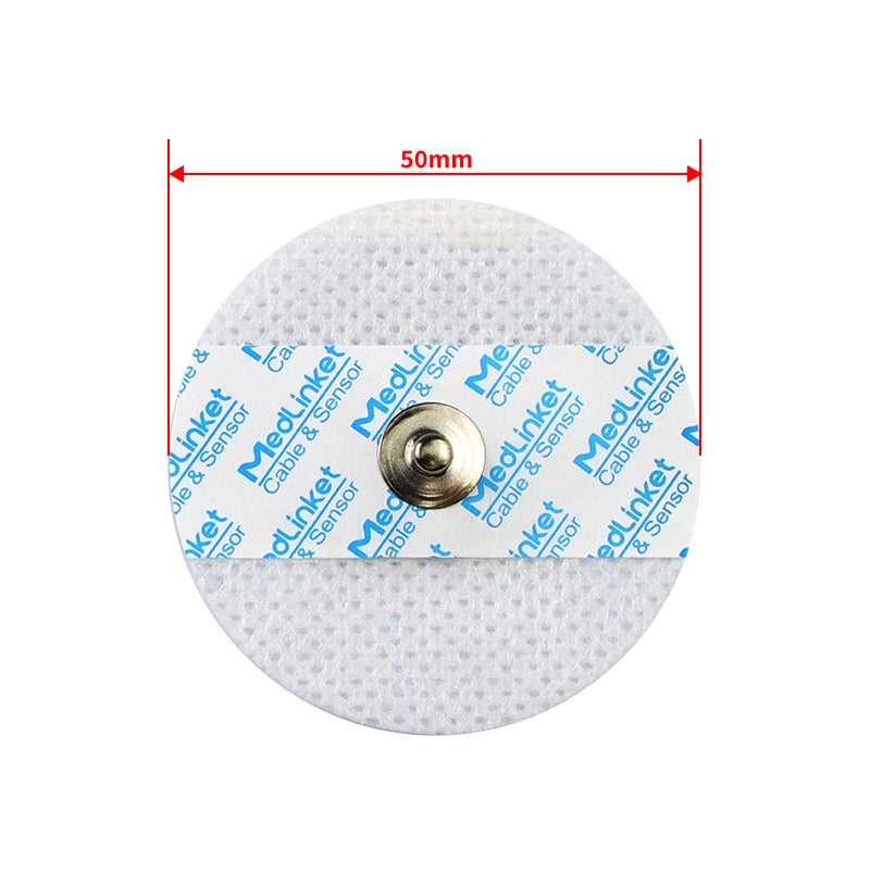
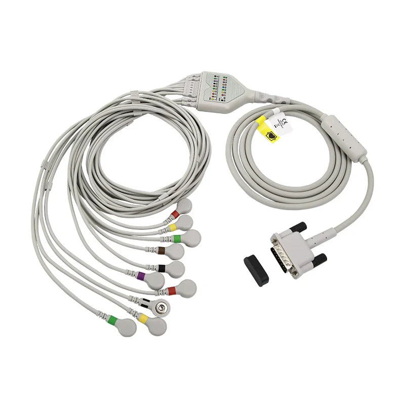
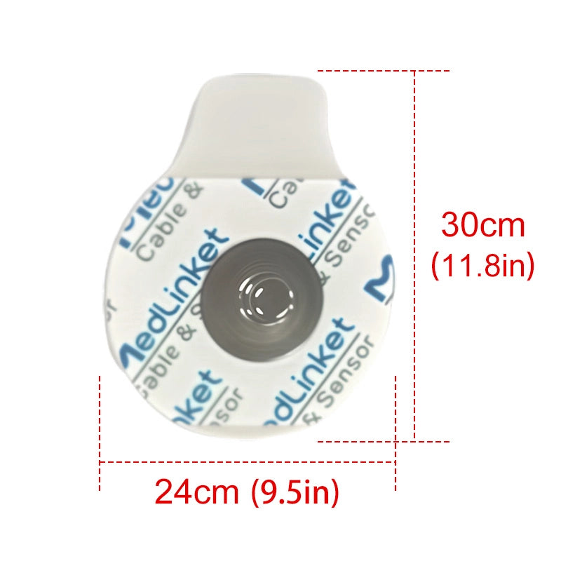
The fix: V4R should be obtained within minutes of identifying inferior STEMI. ST elevation in V4R can resolve as the infarct evolves, so early acquisition is crucial. Keep fresh electrodes and a known-good cable at the bedside so acquisition is never delayed by missing supplies.
Mistake #5: Ignoring Borderline Results
The problem: V4R shows 0.5mm ST elevation, which is dismissed as "not meeting criteria."
The fix: Borderline V4R + hypotension + inferior STEMI = treat as RV involvement. Clinical context matters. When in doubt, avoid preload-reducing agents.
🎯 Keep These at the Bedside So V4R Is Never Delayed
Mistake #4 is waiting too long — fresh electrodes + a 10-lead cable on hand make 30-second V4R acquisition possible every time
Frequently Asked Questions: Right Sided ECG Leads
Q: Do I need to do V4R on every ECG?
A: No. V4R is indicated specifically for inferior STEMI (ST elevation in II, III, aVF) and suspected right ventricular involvement. It's not part of routine 12-lead ECG.
Q: Can I just look at the standard 12-lead for RV MI signs?
A: The standard 12-lead can show clues (ST elevation III > II, ST depression V1-V2), but it cannot reliably detect RV involvement. V4R is required for diagnosis. Don't skip it.
Q: How quickly should V4R be done after identifying inferior STEMI?
A: Immediately. Within minutes of recognizing the inferior STEMI pattern. ST changes in V4R can evolve, and early detection changes management. Don't wait. Keep fresh ECG electrodes ($11.90, 25pcs/bag) at the bedside so a fresh electrode is always within reach.
Q: What if my ECG machine doesn't have a "right sided" setting?
A: Simply move the V4 electrode to the mirror position and run a standard ECG. Manually label the tracing as "V4R." The machine setting is a convenience, not a requirement.
Q: Should I check V4R in NSTEMI?
A: In NSTEMI, V4R is not routinely indicated. However, if there are ischemic changes in inferior leads or clinical suspicion of RV involvement, it's reasonable to check. Use clinical judgment.
Mastering Right Sided ECG Lead Placement
Right sided ECG lead placement is not an advanced technique reserved for specialists—it's a fundamental skill that every clinician managing acute coronary syndromes must master. The 30 seconds it takes to place and record V4R can be the difference between appropriate treatment and catastrophic error.
Remember the key principles of right sided ECG lead placement:
- When: Every inferior STEMI, without exception
- Where: V4R at 5th intercostal space, right midclavicular line (mirror of V4)
- Why: 30-50% of inferior STEMIs involve the RV; treatment is completely different
- What it changes: No nitrates, aggressive fluids, modified cath lab approach
My 68-year-old patient from June 2023 walked out of the hospital because someone took 30 seconds to check V4R. Make sure you do the same for every patient who deserves it.
📚 This Guide is Part of a Series
Return to the main guide: 12 Lead ECG Placement: The Ultimate Guide
Related articles:
Clinical Education Team
Our clinical education team includes emergency medicine and cardiology specialists with extensive experience in acute coronary syndrome management. This guide reflects current AHA/ACC guidelines combined with real-world clinical practice.


