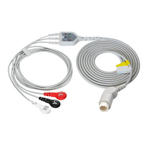
Differences between preductal and postductal oxygen saturation (SpO2) are highly meaningful in neonatal care, especially for detecting critical congenital heart disease (CCHD). Understanding pre- and post-ductal sats—and how they differ—provides clinicians with a non-invasive yet powerful tool to assess newborn circulatory function.
Comparing these two readings helps determine how well-oxygenated blood is before and after it passes the ductus arteriosus, offering vital insight into cardiac shunting and overall cardiopulmonary adaptation. This comparison is essential for early diagnosis, timely intervention, and improved outcomes in vulnerable newborns.

Quick Cultural Note for U.S. Readers
The ductal meaning in neonatology refers to the ductus arteriosus, a normal fetal vessel connecting the pulmonary artery to the descending aorta. It allows most of the right ventricular output to bypass the non-functioning fetal lungs. After birth, as the baby breathes independently, this vessel typically begins closing within hours and fully closes over days. However, abnormal persistence or direction of flow through this duct can signal serious pathology.
I. Pre- and Post-Ductal SpO2: Definitions and Sites
A) Definitions
Pre-ductal SpO2 (right hand): This measures oxygen saturation from the right hand, reflecting systemic arterial blood before it travels past the ductus arteriosus. Because the right subclavian artery branches off the aortic arch proximal to the ductus, the right upper limb provides an excellent approximation of preductal saturation.
Post-ductal SpO2 (foot): Measured on either foot, this reflects oxygen levels after potential mixing of blood via the patent ductus arteriosus into the lower body circulation. Any significant desaturation here compared to the pre-ductal site may indicate right-to-left shunting.
Understanding pre-ductal vs. post-ductal differences enables detection of conditions where deoxygenated blood enters the systemic circulation preferentially in the lower extremities.
| Item | Pre-ductal (Right Hand) | Post-ductal (Foot) |
|---|---|---|
| Physiology | Blood before ductus arteriosus | Blood after potential mixing via PDA |
| Typical Site | Right palm or finger | Dorsum/plantar surface of the foot |
| Clinical Clue | Higher sat vs. foot may suggest differential cyanosis | Lower sat vs. hand may indicate right-to-left shunting |
B) Measurement Sites and Key Steps
Pre-ductal (Right Hand)
- Site: Right palm or finger
- Steps: Clean skin thoroughly; apply appropriately sized pulse oximeter probe snugly without impeding perfusion. Allow 30–60 seconds for stabilization before recording.
Questions about Atrial Septal Defect
byu/PrinceKaladin32 inmedicalschool
Post-ductal (Foot)
- Site: Dorsum or plantar surface of the foot
- Steps: Use wrap-style sensors to ensure consistent contact. Avoid cold extremities or motion artifacts.
Simultaneous measurement at both sites ensures accurate assessment of any gradient.
II. How These Measurements Inform Diagnosis
Assessing pre-ductal and post-ductal sats simultaneously has become standard practice in U.S. hospitals for screening CCHD. Many infants with structural heart defects appear clinically well at birth, making routine physical exams insufficient.
A key diagnostic clue lies in comparing values:
- Normal finding: Both readings ≥95%, with a difference ≤2%.
- Suspicious finding: Difference >2% between preductal vs. postductal coarctation-related patterns or other cyanotic lesions.
- High-risk threshold: ≥4% difference triggers immediate echocardiographic evaluation.
For example, in preductal vs. post-ductal comparisons, if the foot (post-ductal) reading is significantly lower than the right hand (pre-ductal), it suggests that poorly oxygenated blood is shunting from the pulmonary artery into the descending aorta via a patent ductus arteriosus—a hallmark of right-to-left shunt physiology.
U.S. guidelines (endorsed by the American Academy of Pediatrics) recommend universal screening using pre- and post-ductal sats between 24–48 hours of life. A pass is defined as SpO2 ≥95% in both limbs with ≤3% difference. Values below 90% constitute automatic failure, warranting urgent investigation.
III. Clinical Meaning of SpO2 Differences
A) Normal Ranges Across Populations
| Population | Typical SpO2 Range |
|---|---|
| Healthy adults | 95%–100% |
| Newborns | Gradually rise during first 24 hours; target 90%–97% |
| High altitude residents | ~90%–95% |
| COPD patients | Often 88%–92% (medical supervision) |
| Pulmonary hypertension | Maintain >92% to reduce right heart strain |
Persistent SpO2 <90% in a term infant raises concern for hypoxemia regardless of site.
B) Interpreting Pre- vs Post-Ductal Differences
- ≤2% difference: Indicates minimal shunting across the ductus — expected in healthy transition.
- >2% difference: Raises suspicion for right-to-left shunting due to congenital anomalies such as tetralogy of Fallot, transposition of the great arteries, or persistent pulmonary hypertension of the newborn (PPHN).
- ≥4% difference: Strongly suggestive of CCHD; requires prompt echocardiography.
Other causes include:
- PPHN: Elevated pulmonary vascular resistance leads to right-to-left ductal shunting.
- Coarctation of the aorta: Often presents with lower blood pressure and reduced saturation in the legs, detectable only when pre- and post-ductal sats are compared.
This distinction makes preductal vs. postductal monitoring indispensable in differentiating central cardiovascular pathology from primary respiratory disease.

IV. What to Do When the SpO2 Difference Exceeds 2%
A) Initial Assessment
- Confirm correct placement: Right hand = preductal, foot = post-ductal.
- Rule out technical errors: Poor probe contact, nail polish, cold extremities, movement.
- Correlate clinically: Assess for tachypnea, retractions, murmurs, cyanosis, or poor perfusion.
B) Further Testing
- Echocardiography: Gold standard for evaluating anatomy and shunt direction (e.g., Qp:Qs ratio)
- Arterial blood gas (ABG): Assesses true PaO2, acid-base status
- Chest X-ray/CT: Evaluates lung fields, heart size, and possible aortic abnormalities like rib notching in chronic coarctation
C) Interventions Based on Cause
- Right-to-left shunt: May require prostaglandin E₁ infusion to maintain ductal patency until surgery (e.g., duct-dependent lesions)
- PPHN: Inhaled nitric oxide, high-frequency ventilation, or ECMO
- Coarctation of the aorta: Surgical or catheter-based repair; monitor for post-op hypertension
Early recognition using pre-ductal and post-ductal sats can prevent shock, acidosis, and end-organ damage.
V. Standardized Newborn Screening: Timing & Workflow
A) Optimal Timing and Technique
- When: Screen between 4–12 hours after birth, ideally before nursery discharge.
- How: Place probes simultaneously on the right hand (pre-ductal) and one foot (post-ductal) to capture real-time differential.
Accurate technique directly impacts reliability—errors here could miss life-threatening conditions.
B) Continuous Monitoring & Documentation
- Set alarm limits: Low at 89%, high at 96%
- Record all values, interventions, and rechecks
- Trend data rather than relying on single measurements
C) Managing Abnormal Results
1) Rule Out Artifacts First
Ensure no sensor slippage, ambient light interference, or vasoconstriction. Recheck after warming or calming the infant.
2) If Infant Is Unstable – Treat First
Red flags:
- Respiratory rate >60/min
- Central cyanosis
- Bradycardia (<100 bpm) or tachycardia (>220 bpm)
- Capillary refill >3 seconds
Immediate actions:
- Administer supplemental O2 cautiously
- Begin cardiorespiratory monitoring
- Prepare for NICU transfer
3) Focused Workup
- Repeat pre- and post-ductal sats in ~30 minutes
- Perform ABG and chest X-ray
- Schedule urgent echocardiogram if difference persists ≥4%
Suspected preductal vs. postductal coarctation? Compare arm and leg blood pressures—classic sign is higher BP in arms.
Additional tests:
- BNP: Evaluate for heart failure
- Photoplethysmography (PPG): Assess peripheral perfusion trends
- High-resolution CT: For suspected vascular malformations or complex anatomy
4) Transfer & Multidisciplinary Care
If diagnosis remains unclear or intervention is needed, expedite transfer to a pediatric cardiac center. Collaborative teams involving cardiology, pulmonology, and neonatology optimize outcomes.
Follow-up:
- Serial echos every 3–6 months for confirmed CCHD
- Parent education on warning signs, feeding challenges, infection prevention
- Long-term O2 users: Regular ABGs and pulmonary function testing
5) Simplified Process Map
Abnormal SpO₂ → Check for artifacts
→ Signs of instability? → Stabilize + Transfer
→ No emergency? → Repeat SpO₂ + CXR + ABG
→ Difference still >2%? → Echocardiogram
→ Diagnosis? → Treat / Refer → Follow-up
→ No clear cause? → Advanced imaging (CT), BNP, PPG
6) Key Principles
- Trend over time matters more than isolated values
- Individualize targets based on gestational age, comorbidities, and clinical context
- Embrace team-based care—cardiac and pulmonary systems are deeply intertwined
VI. Special Considerations
A) Preterm vs Term Infants
- Target SpO2: 91%–95% for preterms; similar for term infants unless respiratory distress syndrome or PDA complicates care.
- Lower thresholds accepted under controlled settings to avoid retinopathy of prematurity.
B) Cyanotic Heart Disease
In known cases, cardiology may set individualized goals (e.g., 75%–85%) to balance oxygen delivery and pulmonary overcirculation risk.

Final Thoughts
Accurate measurement of pre- and post-ductal sats is a cornerstone of modern neonatal screening. By understanding preductal vs. postductal physiology—and recognizing what constitutes a concerning gap—clinicians can identify silent but deadly conditions like critical congenital heart disease long before symptoms emerge.
Whether you're interpreting subtle gradients in a vigorous newborn or managing a critically ill infant with suspected preductal vs. postductal coarctation, this simple, non-invasive test offers profound clinical value. When combined with standardized protocols, artifact awareness, and multidisciplinary follow-up, pre-ductal and post-ductal sats remain one of the most effective tools in preventing delayed diagnoses and improving survival rates.
FAQ:
Q1:
Why is preductal higher than postductal?
In certain congenital heart defects—such as transposition of the great arteries (TGA) or critical coarctation of the aorta—well-oxygenated blood is preferentially directed to the upper body, while blood reaching the lower body may mix with deoxygenated blood through a patent ductus arteriosus. As a result, the right hand (preductal) saturation is often higher than the foot (postductal) saturation. This pattern, known as differential cyanosis, is clinically meaningful and helps flag ductal-dependent or right-to-left shunting physiology.
Q2:
Why do we perform pre-ductal oxygen saturations on babies?
Because the right hand receives blood from the brachiocephalic (innominate) artery before the ductus arteriosus, it provides the best snapshot of central systemic oxygenation. Comparing right-hand (preductal) and foot (postductal) values helps detect differential cyanosis, supports early identification of critical congenital heart disease (CCHD), and guides decisions about urgent imaging or transfer—often starting with an echocardiogram.
Q3:
Why is the right hand used for pre-ductal pulse oximetry monitoring?
Anatomically, the right subclavian artery branches from the aorta proximal to the ductus arteriosus, so the right hand reflects preductal blood. The left subclavian artery arises distal to the ductus and may already contain mixed (postductal) blood. Standardizing preductal measurements on the right hand improves consistency and diagnostic accuracy across care teams and facilities.
As experts in patient monitoring, MedLinket designs and manufactures high-performance patient monitoring accessories trusted in more than 120 countries and by 2.000+ hospitals worldwide.
With over 20 years of engineering experience, an ISO 13485–based quality system, and major market certifications (including FDA and CE), our solutions deliver reliable accuracy and seamless compatibility across leading platforms such as Philips, GE, Mindray, Dräger, Masimo, and Nellcor.
To learn more about pulse oximeters, NICU/ICU monitoring, and fetal/neonatal measurement solutions, browse our full range of Disposable SpO2 Sensors—from ECG trunk cables to direct-connect EKG cables—or email our experts at shopify@medlinket.com today.


