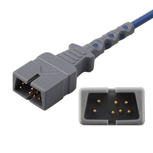
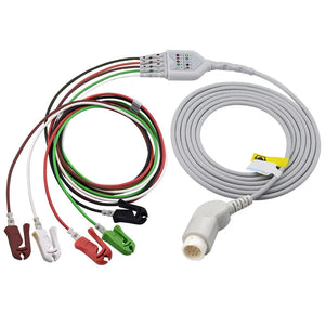
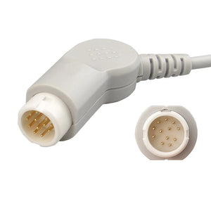
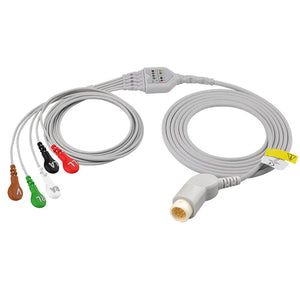
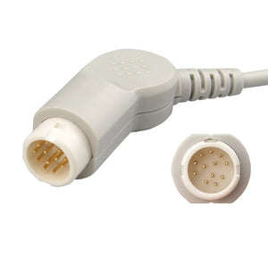
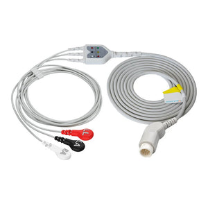
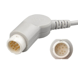
For routine veterinary ECG monitoring, place the limb electrodes near the limbs and confirm the cable standard before connecting the patient. In the common AHA scheme, RA is white on the right forelimb, LA is black on the left forelimb, LL is red on the left hindlimb, and RL is green on the right hindlimb. A 5-electrode cable also includes a V/precordial electrode whose position should follow the monitor instructions and veterinarian-directed protocol. Improve contact by parting or clipping fur and using an approved conductive medium.
An ECG is only as trustworthy as its lead placement. Put the electrodes in the wrong spot, or get poor skin contact through fur, and the trace is noisy or misleading before anyone tries to read it. Veterinary ECG placement differs from the human chest-lead setup most people picture — on dogs and cats the leads go on the limbs — and the fur adds a contact challenge humans don't have. This guide covers the standard lead positions, the color convention, and how to get a clean trace on a furred patient.
ECG is one of the core parameters in small-animal anesthesia monitoring. Veterinary monitoring placement differs from human chest-lead placement because the anatomy, coat, and electrode sites are different. Species-specific interpretation—what a tracing means for an individual animal—always rests with the attending veterinarian; this guide focuses on obtaining a stable monitoring signal.
On this page
Veterinary monitoring uses animal-specific positionsDogs and cats commonly use limb electrodes near the elbow and stifle regions for rate-and-rhythm monitoring. A 5-electrode cable also adds a V/precordial electrode according to the monitor and clinical protocol. This differs from the standard human precordial layout and from a veterinarian-directed diagnostic ECG.
The standard limb-lead color convention
Veterinary monitoring systems commonly use either AHA or IEC lead labeling. The table below shows the usual limb assignments and the additional V/precordial electrode found on a 5-electrode cable. Always confirm the printed lead labels and the monitor instructions before use.
| Electrode | Usual location | AHA color | IEC color |
|---|---|---|---|
| RA | Right forelimb | White | Red |
| LA | Left forelimb | Black | Yellow |
| LL | Left hindlimb | Red | Green |
| RL | Right hindlimb / reference | Green | Black |
| V / C | Precordial monitoring position selected by protocol | Brown | White |
Color schemes differ—check the cable labelsAHA and IEC use different colors for the same electrode labels. Match RA, LA, LL, RL, and V/C by the printed cable label and monitor instructions—not by color memory alone. The V/C electrode on a 5-electrode cable is not interchangeable with one of the four limb electrodes.
Where the leads go on a dog or cat
The illustration shows the four limb-electrode positions for a monitoring ECG on a dog in lateral recumbency; the same placement logic applies to cats.

Electrodes attach near the elbow and stifle regions rather than on the paws. A 5-electrode system adds a V/precordial electrode placed according to the monitor instructions and the attending veterinarian’s protocol.
3-lead vs 5-lead: which to use
Cable configuration is selected according to the monitoring goal. Fewer electrodes simplify rapid rate-and-rhythm monitoring, while a 5-electrode cable adds another monitoring vector. A diagnostic 12-lead ECG is a separate veterinarian-directed examination.
| Configuration | Typical setting | Why |
|---|---|---|
| 3-electrode monitoring | Emergency, transport, recovery, or basic rate-and-rhythm monitoring | RA, LA, and LL provide a rapid monitoring configuration with fewer wires |
| 5-electrode monitoring | Operating room, ICU, anesthesia, or extended monitoring | Adds RL reference and a V/precordial electrode for another monitoring vector |
| Diagnostic 12-lead ECG | Cardiac diagnostic evaluation | A separate standardized acquisition and interpretation workflow led by a veterinarian |
Getting a clean trace on a furred patient
Fur, patient motion, and cable pull make skin contact especially important in dogs and cats. Whatever electrode style is used, the goal is a stable connection to prepared skin without excessive pressure or tissue injury.
- Part or clip the fur. Get the contact point down to skin — part the coat for clips, or clip a small patch for adhesive electrodes on a dense coat.
- Use an approved conductive medium. Apply ECG gel or another product permitted by the electrode and clinic protocol. Avoid pooling flammable alcohol when electrosurgery or other ignition sources are present.
- Use clips where appropriate. Atraumatic clips at the limbs are quick for monitoring; adhesive electrodes suit clipped skin and longer runs.
- Manage cable tension. Support and route the cable so movement does not rotate, lift, or detach the electrode. Strain relief is especially important during transport and repositioning.
- Keep the patient still and warm. Shivering and movement both add artifact; a calm, warm patient gives a cleaner trace.
ECG electrodes and leads for veterinary use
Veterinary ECG accessories must maintain contact through fur and movement while matching the monitor connector, lead count, and AHA or IEC standard.

MedLinket supplies veterinary ECG electrodes, lead wires, and patient cables for compatible monitoring systems.
Explore the full range in Veterinary Supplies. ECG is one layer of a monitoring setup — for the whole picture see anesthetic monitoring in small animals, and to stock a mixed clinic see building a veterinary monitoring accessory kit.
Why source veterinary ECG accessories from MedLinket
MedLinket’s ECG product range includes electrodes, lead wires, patient cables, telemetry cables, and related monitoring accessories.
- A full in-house ECG line. MedLinket (Shenzhen Med-link Electronics Tech Co., Ltd.) manufactures its own ECG electrodes, lead wires, main cables, telemetry and Holter cables, and related consumables — ECG and electrosurgery is one of its core product lines, and animal healthcare is one of four core clinical solution areas.
- Engineered electrode options. MedLinket product materials describe eccentric electrode structures intended to move cable pull away from the conductive area and permit lead rotation. Confirm the exact product design and supporting documentation before ordering.
- Broad, honestly labeled compatibility. ECG leads are available in AHA and IEC standards and are designed for mainstream monitor technologies. MedLinket is an independent third-party manufacturer; "compatible with" never implies OEM manufacture or brand endorsement.
- Product-level documentation. Quality-system, registration, and certification claims should be checked for the exact product, configuration, and destination market rather than generalized across the full catalog.
Learn more about MedLinket and review the company’s certification information. Product-level certifications and compatibility should be confirmed for the exact SKU and destination market.

Frequently asked questions
Where do ECG leads go on a dog?
How is veterinary ECG placement different from human?
How do you get a good ECG trace through fur?
Should I use a 3-lead or 5-lead ECG?
What do AHA and IEC color codes mean?
This article provides general educational and product-selection information. It is not a substitute for a veterinarian’s examination, diagnostic ECG protocol, clinical interpretation, or the device instructions for use. Lead labels and colors vary by cable standard; always confirm the monitor, connector, electrode count, printed labels, electrode position, and intended use. Product compatibility and certification can vary by SKU, configuration, and market. Third-party brand references indicate compatibility only and do not imply OEM manufacture, authorization, affiliation, or endorsement.