Small-animal anesthesia monitoring combines five electronic parameter groups: SpO₂ for oxygenation, EtCO₂ for ventilation, ECG and heart rate for rhythm, blood pressure for circulation, and temperature. A dedicated anesthetist must also assess anesthetic depth, airway, breathing, pulses, mucous membranes, pain, and trends.
Anesthetizing a dog or cat is a continuum that begins with patient assessment and equipment preparation and continues through induction, maintenance, extubation, and recovery. A veterinary anesthesia monitor can display several parameters at once, but the display is only one part of the safety system. The trained person monitoring the patient must interpret the values, inspect the patient and equipment, recognize trends, and respond to change.
The 2025 American College of Veterinary Anesthesia and Analgesia guidelines organize monitoring around circulation, oxygenation, ventilation, body temperature, anesthetic depth, neuromuscular blockade when used, personnel, and record keeping. This article translates that framework into the five electronic parameter groups most clinics associate with veterinary monitoring equipment, while preserving the hands-on assessment that no multiparameter vet monitor can replace.
The monitor is not the anesthetist Electronic monitoring and hands-on assessment should be used together. A stable number can be wrong because of motion, poor probe contact, an incorrect cuff, circuit leaks, an electrical artifact, or a delayed display. A trained person should remain with the anesthetized patient, assess depth and clinical signs, document values, and intervene according to the veterinarian-approved plan.
The five key anesthesia monitoring parameters
| Parameter | Primary question | Typical accessory | Major limitation |
|---|---|---|---|
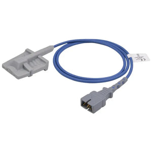
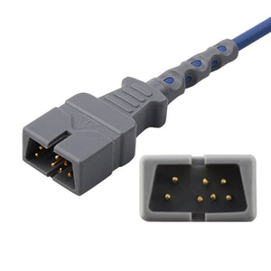
| SpO₂ | Is hemoglobin adequately oxygenated? | Veterinary tongue clip or multi-site SpO₂ sensor | Does not measure ventilation; motion, low perfusion, site, and sensor fit affect reliability |
| EtCO₂ | Is the patient ventilating, and is the airway/circuit connected? | Mainstream sensor and airway adapter, or sidestream sampling line | Leaks, dead space, sampling dilution, obstruction, and nonintubated interfaces affect values |
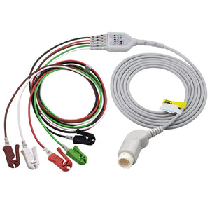
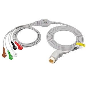
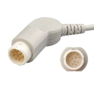
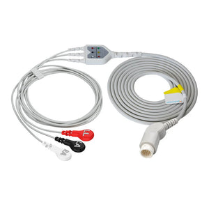
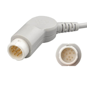
| ECG / heart rate | What is the electrical rate and rhythm? | Patient cable, lead wires, clips, needles, or electrodes | Electrical activity does not guarantee an effective pulse or adequate blood pressure |
| Blood pressure | Is arterial pressure adequate for the patient and procedure? | Measured NIBP cuff and hose, Doppler, oscillometric monitor, or IBP system | Cuff size, site, heart level, motion, algorithm, and method affect interpretation |
| Temperature | Is the patient losing or gaining heat? | Rectal or esophageal temperature probe plus approved warming equipment | Site lag, shallow placement, poor contact, and unsafe heating methods can mislead or injure |
How often should values be recorded?
The 2025 ACVAA guidelines state that heart rate, arterial blood pressure, arterial oxygen saturation, and end-tidal carbon dioxide should generally be recorded every five minutes. Other physiologic variables should be recorded at least every 15 minutes, while patient observation remains continuous. Unstable patients may require more frequent measurement and documentation.
Assess airway, breathing effort, pulse quality, mucous membranes, anesthetic depth, eye position, jaw tone, reflexes, surgical stimulation, pain, and equipment function.
Record trends and interventions, not only alarm events. More frequent recording can be appropriate when the patient is unstable.
The 2025 guideline specifically recommends measuring rectal temperature at least every 15 minutes in moderately to heavily sedated or anesthetized small animals.
Document drugs, monitored values, patient position, equipment, events, troubleshooting, interventions, and recovery.
Follow the clinic's current protocol These intervals summarize the 2025 ACVAA guideline and are not a substitute for local regulation, equipment instructions, patient-specific needs, or the veterinarian-approved anesthetic plan.
The anesthetized patient at a glance
1. SpO₂: monitor oxygenation and signal quality
Pulse oximetry estimates arterial hemoglobin oxygen saturation. It does not show whether carbon dioxide is being eliminated, so a normal SpO₂ value cannot rule out hypoventilation, especially when supplemental oxygen is being delivered.
- In anesthetized dogs and cats, the tongue is often a practical transmission site, but the best site still depends on the probe, patient, perfusion, and procedure.
- Confirm a stable pleth waveform and compare the pulse-oximeter pulse rate with ECG, auscultation, palpation, or an arterial pressure trace.
- Reassess motion, probe pressure, optical alignment, tissue thickness, ambient light, hypothermia, hypotension, and compatibility when the reading is unstable.
Use the dog and cat pulse oximeter placement guide and veterinary SpO₂ troubleshooting guide for the full workflow.
2. EtCO₂: monitor ventilation, airway, and circuit function
Capnography measures carbon dioxide at the end of exhalation. In an intubated patient, it helps confirm continuing airway connection and provides information about ventilation, circulation, rebreathing, obstruction, disconnection, and breathing-circuit function. In a sedated or nonintubated patient, nasal, oral, mask, or other sampling interfaces may be used, but leaks and room-air dilution can make the number less representative.
- Mainstream: the sensor is placed at the airway; consider added weight and dead space for very small patients.
- Sidestream: gas is drawn through a sampling line; check delay, blockage, moisture, leaks, water traps, and sampling flow.
- Waveform matters: interpret the capnogram shape, respiratory rate, trend, and patient condition—not only a single EtCO₂ number.
Browse EtCO₂ equipment, EtCO₂ sensors, and disposable EtCO₂ sampling lines.
3. ECG and heart rate: electrical rhythm is not the pulse
ECG shows cardiac electrical activity and helps identify rate and rhythm changes. It does not directly prove that each electrical complex produces effective circulation. Compare ECG heart rate with the pulse-oximeter rate, arterial pressure waveform, Doppler signal, auscultation, or palpated pulse. A discrepancy can indicate artifact, pulse deficit, or hemodynamically important arrhythmia.
- Prepare the contact site according to clinic protocol and minimize cable movement.
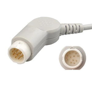
- Match 3- or 5-lead configuration, connector, and AHA or IEC color standard to the monitor.
- Recheck electrode contact, lead placement, grounding, electrical interference, and patient position when the trace is noisy.
Read the veterinary ECG lead-placement guide , then browse ECG cables and lead wires and disposable ECG electrodes.
4. Blood pressure: cuff, site, and method must stay consistent
Indirect blood pressure can be measured with oscillometric or Doppler methods. For a dog or cat NIBP cuff, measure the selected limb or tail circumference and choose a compatible cuff whose width is approximately 30%–40% of that circumference. Keep the measurement site near heart level and use the same method and site when trending values.
- A cuff that is too narrow tends to overestimate pressure.
- A cuff that is too wide tends to underestimate pressure.
- Motion, low perfusion, hose leaks, connector mismatch, and algorithm limitations can cause failed or implausible cycles.
- For critical patients, direct arterial blood pressure may be selected by the veterinarian.
See the dog and cat blood pressure cuff guide and browse veterinary blood pressure monitors.
5. Temperature: measure early and warm safely
Hypothermia can delay drug metabolism and recovery and contribute to impaired perfusion, cardiovascular dysfunction, respiratory compromise, and patient discomfort. Temperature should be measured rather than estimated, heat loss should be limited early, and warming should continue into recovery when indicated.
- Rectal and esophageal temperatures are clinically reasonable sites, but their response times and placement requirements differ.
- Match the temperature probe's thermistor family, connector, site, cable length, and instructions to the monitor.
- Use warming devices intended for anesthetized patients. Improvised or unregulated heat sources can cause severe thermal injury.
- Inspect the skin and contact areas and avoid placing an anesthetized patient directly against an uncontrolled heat source.
Browse temperature monitoring products, reusable temperature probes, and disposable warming blankets.
Anesthetic depth and hands-on assessment
Anesthetic depth is not one of the five electronic values, but it is a minimum clinical responsibility. A dedicated anesthetist repeatedly evaluates eye position, jaw and body tone, palpebral or withdrawal reflexes where appropriate, response to stimulation, respiratory pattern, pulse quality, mucous membranes, analgesia, and patient positioning.
Trend relationships, not isolated numbers A rising heart rate with hypertension can mean something different from a rising heart rate with falling blood pressure. A low SpO₂ value with a clean pleth waveform deserves a different response from a low value with a pulse-rate mismatch. Interpret the whole patient, waveform quality, recent interventions, anesthetic depth, and the direction of change.
Monitoring through the four anesthetic phases
Preanesthesia
Assess and stabilize the patient, create an individualized plan, select correct accessory sizes, check equipment, establish baseline values, and prepare warming.
Induction
Apply ECG, SpO₂, and BP monitoring promptly; secure the airway as planned and begin capnography when the selected interface permits.
Maintenance
Maintain continuous observation, record values, assess depth and analgesia, prevent heat loss, and respond to trends in ventilation, oxygenation, circulation, and temperature.
Recovery
Continue monitoring through extubation and until the patient is appropriately alert, warm, ventilating, oxygenating, and stable for the next level of care.
Recovery remains a high-risk period AAHA guidance reports that a large proportion of anesthetic-related dog and cat deaths occur postoperatively, with many occurring during the first three hours. Recovery should receive the same vigilance as maintenance, including respiratory rate, SpO₂, heart rate, blood pressure when indicated, temperature, airway protection, pain, and warming.
MedLinket veterinary monitors and accessory families
MedLinket offers dedicated animal-use devices and five veterinary accessory families. Veterinary and human product specifications should not be combined unless the exact SKU documentation supports the intended use.
| Product or family | Anchored facts | Role in the anesthesia setup |
|---|---|---|
| ESM601 | Veterinary monitor with 5.5-inch touch screen; unit weight under 0.5 kg; SpO₂, TEMP, NIBP, HR, and EtCO₂ | Compact multiparameter host for animal monitoring |
| AM-806VB-E | Veterinary SpO₂/temperature device with Bluetooth; anchor ranges include SpO₂ 35%–100%, pulse rate 30–300 bpm, and temperature 25–45°C | Portable oxygenation, pulse, and temperature monitoring |
| ESM303 | Veterinary oscillometric blood pressure monitor; current product page lists five cuff ranges covering 3–15 cm | Dedicated animal NIBP system |
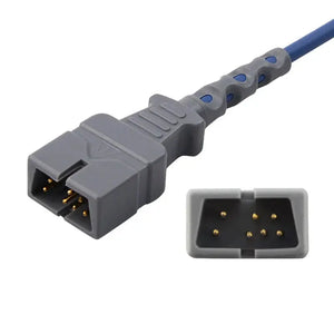
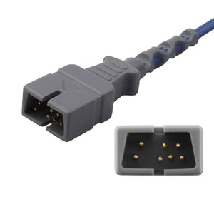
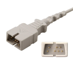
| S series | Veterinary SpO₂ examples include H tongue clip and specialized R1/R2 rectal styles | Match sensor site, oximetry technology, connector, and host device |
| E series | Veterinary ECG accessory examples include EA020S5A and V0015A/B | Match ECG lead count, connector, termination, and AHA/IEC standard |
| T series | Temperature examples include W0024D for AM-806VB and W0001B compatible with Philips A3 | Match thermistor family, connector, measurement site, and cleaning method |
| N series | Five veterinary NIBP cuff sizes in the anchor cover labeled circumferences from 3–15 cm | Measure the patient and match cuff, hose, connector, and monitor |
| C series | Mainstream CA10-001/002 and sidestream CA20-001/003/004/005 examples | Match capnography method, airway interface, consumables, and host connection |
Veterinary monitoring equipment and accessory links
Compatibility information needed Provide the monitor manufacturer and full model, parameter module, current accessory part number, connector photos, patient species and size, intended site, cable length, AHA or IEC ECG standard, NIBP circumference range, reusable or single-patient preference, and required quantity. Third-party brand references indicate compatibility only, not OEM manufacture, authorization, affiliation, or endorsement.
Why source veterinary monitoring accessories from MedLinket?
- Dedicated veterinary structure. MedLinket separates animal-use devices and S/E/T/N/C accessory families from human device specifications.
- Five-parameter accessory coverage. MedLinket's veterinary product system includes SpO₂, ECG, temperature, NIBP, and EtCO₂ accessory families plus veterinary monitoring devices.
- Compatibility-first selection. Host technology, connector, patient size, site, cable, and clinical workflow should be checked before a product is represented as compatible.
- Product-level claims. Specifications, certification, validation, warranty, and intended-use statements should remain tied to the exact SKU and current documentation.
Learn more on the MedLinket company page, review certifications, or use the contact page for a model-level compatibility check.
Frequently asked questions
What parameters should be monitored during small-animal anesthesia?
Is EtCO2 only needed for intubated dogs and cats?
How often should anesthesia monitoring values be recorded?
Why is temperature monitoring important during veterinary anesthesia?
Can a multiparameter veterinary monitor replace a dedicated anesthetist?
What is the difference between SpO2 and EtCO2?
What veterinary blood pressure cuff size should be used during anesthesia?
What information is needed to order veterinary monitoring accessories?
This article provides general educational and product-selection information. It is not a substitute for a veterinarian-approved anesthetic plan, a trained anesthetist, current professional guidelines, equipment instructions, local regulations, or clinical judgment. Monitoring needs and target values depend on species, patient status, procedure, drugs, equipment, and spectrum of care. Confirm the exact product specification, intended use, certification, connector, and monitor compatibility before purchase or use.