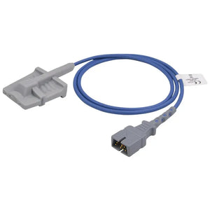
An inaccurate or missing veterinary SpO₂ reading is often caused by weak perfusion, motion, poor probe alignment, unsuitable tissue thickness, fur, ambient light, or sensor incompatibility. Check the animal first. Then confirm a stable pleth waveform, match pulse rate to an independent source, reposition the probe, reduce interference, and test the hardware only after the application is sound.
A veterinary pulse oximeter may show dashes, a noisy waveform, delayed updates, or a saturation value that does not fit the animal's clinical condition. The correct response is not to assume that the sensor is broken—or that the displayed value is harmless artifact. Pulse oximetry needs a detectable arterial pulse signal and a clean optical path between the light emitter and detector. Anything that disrupts either one can produce dropout or bias.
This article covers the animal-specific layer: fur, small or irregular anatomy, pigmentation, motion, low peripheral perfusion, probe pressure, and veterinary sensor fit. For choosing the initial site, start with veterinary pulse oximeter sensor placement . The shared physics and human-use limitations are summarized in how SpO₂ sensors work and SpO₂ sensor troubleshooting . Keeping the animal-specific troubleshooting here reduces overlap between the veterinary and news content clusters.
Assess the patient before the sensor A low, absent, or unstable reading can reflect measurement failure, but it can also accompany true hypoxemia, poor circulation, hypotension, hypothermia, arrhythmia, or clinical deterioration. Evaluate airway, breathing, circulation, mucous membranes, pulse quality, heart rate, ventilation, and the attending veterinarian's assessment. Do not delay patient care while troubleshooting a monitor.
The 60-second veterinary SpO₂ troubleshooting sequence
Work from signal validity and patient condition toward placement, interference, compatibility, and hardware. The order prevents unnecessary probe replacement and reduces the risk of dismissing a real clinical problem.
Check the animal and the independent pulse
Confirm the patient's clinical status and compare the oximeter pulse rate with ECG heart rate, auscultation, or a palpable pulse. A large mismatch suggests motion, weak signal, arrhythmia-related detection problems, or poor application.
Patient stable and pulse agrees → review waveform quality. Patient abnormal or pulse disagrees → reassess clinically and continue troubleshooting.
Inspect the pleth waveform and signal indicator
Look for a repeating waveform with consistent timing and amplitude. Perfusion Index or another signal-quality indicator can help, but its scale and useful range are monitor-specific. Do not use one numerical PI cutoff as a universal proof that every veterinary reading is accurate.
Stable trace and matching pulse → interpret with the patient. Flat, irregular, or motion-dominated trace → correct the site and application.
Reposition and realign the emitter and detector
The light path must cross suitable pulsatile tissue. Make sure the two optical elements face each other, the probe is not rotated, tissue is not folded unpredictably, and the clip is neither loose nor excessively tight. Excessive pressure can reduce local blood flow.
Signal recovers → secure the probe without constricting tissue. Still poor → select another anatomically suitable site.
Choose a site that fits the species, patient, and probe
There is no universal “tongue > ear > toe” ranking for every dog, cat, state of consciousness, probe design, or monitor. Select thin tissue that fits the sensor, has adequate local perfusion, allows optical alignment, and can be kept still. In anesthetized dogs, the tongue is often practical; in cats or awake patients, another site may fit better.
Appropriate site works → document the site and probe style. Still unstable → remove interference and address perfusion.
Reduce motion, fur, debris, and ambient light
Stabilize the sensor and cable, minimize patient motion with appropriate handling, remove dried secretions or debris, and shield the probe from strong surgical, warming, or sunlight sources. When fur prevents contact or alignment, choose exposed tissue or carefully clip only the small application area when appropriate.
Waveform improves → continue monitoring. Still poor → reassess temperature, circulation, and device compatibility.
Address low perfusion and hypothermia
Cooling, vasoconstriction, hypotension, shock, and low cardiac output can reduce pulsatile flow at a peripheral site. Reassess the patient, follow the veterinarian's warming and circulatory-management plan, and consider a better-perfused site that fits the sensor.
Signal returns as circulation improves → keep trending. No improvement → check compatibility and hardware.
Inspect compatibility, cable, connector, and sensor
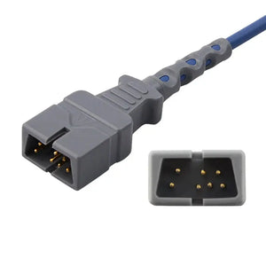
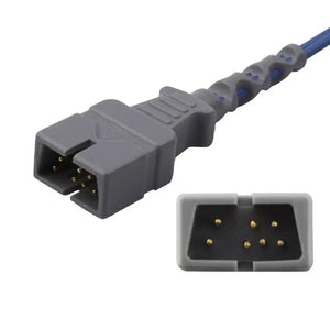
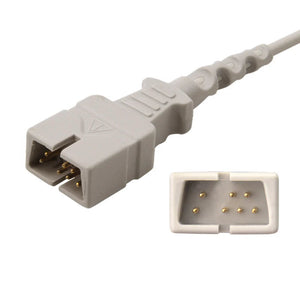
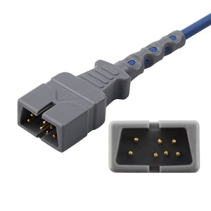
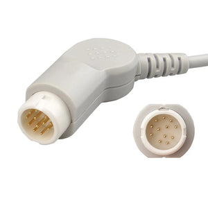
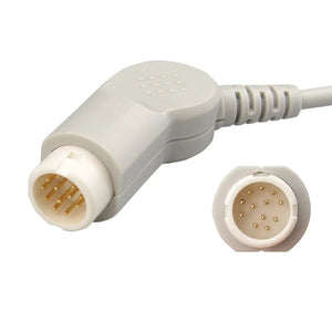
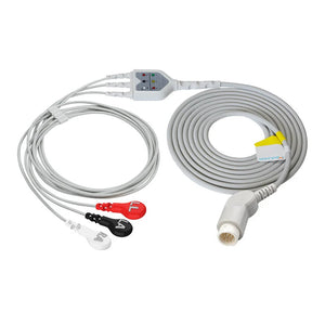
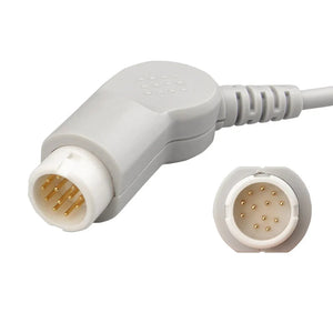
Confirm the monitor's oximetry technology, connector, sensor type, and intended site. Inspect the emitter, detector, clip, cable strain relief, connector pins, and extension cable.

Test a known-good compatible sensor and cable when the patient and application have been addressed.
The site-swap test Apply the same sensor correctly to a second appropriate site on the same stable patient. If the waveform and pulse agreement improve, the original site, tissue fit, pressure, or application was the likely cause. Persistent failure does not prove that the sensor is defective; next test a known-good compatible sensor, cable, and monitor connection.
Common causes of inaccurate veterinary SpO₂ readings
| What you see | Likely contributors | First checks |
|---|---|---|
| No value or repeated dropout | Low local perfusion, poor contact, unsuitable tissue thickness, cable movement, incompatibility | Check patient and pulse; realign probe; change to a fitting site; inspect connector |
| Jumping SpO₂ and pulse rate | Patient motion, shivering, panting-related movement, cable pull, loose sensor | Compare with ECG/palpated pulse; stabilize patient, sensor, and cable |
| Stable number but poor or implausible waveform | Artifact, venous pulsation, periodic motion, optical shunting, delayed display | Do not trust the number alone; reposition and verify pulse agreement |
| Signal worsens during anesthesia | Cooling, vasoconstriction, hypotension, probe displacement, pressure on tissue | Reassess temperature and circulation; inspect site pressure and alignment |
| Works on one site but not another | Anatomy, pigmentation, fur, tissue thickness, perfusion, sensor fit | Use the site that provides correct optical alignment and stable pulse signal |
| Fails with every appropriate application | Damaged sensor or cable, connector fault, extension cable, wrong oximetry technology | Test a known-good compatible sensor/cable and confirm monitor technology |
Animal-specific causes in detail
Probe alignment and tissue thickness
Pulse oximetry depends on red and near-infrared light passing through pulsatile tissue. If the emitter and detector are offset, if the tissue is too thin or too thick for the probe geometry, or if light reaches the detector without crossing an arterial bed, the monitor may display no value or an implausible one. This is why the same sensor can work on a tongue but fail on a narrow lip, pinna, skin fold, or very small toe.
Fur, debris, and moisture
Fur can interfere with probe contact, fixation, and optical alignment, especially on a paw, toe web, limb fold, or tail. Dirt, dried blood, saliva buildup, gel residue, and other debris can also affect contact or allow the sensor to slip. Clean and dry the selected area according to clinic protocol. Prefer a naturally exposed site; clip only a small area when necessary and clinically appropriate.
Motion and cable pull
Struggling, shivering, tremor, chewing, panting-related tissue movement, and a wagging tail can introduce periodic or irregular artifact. Cable weight can rotate a clip even when the animal is still. Route and secure the cable so it does not pull on the probe, select a less mobile site, and use gentle handling or veterinarian-directed sedation only when clinically indicated. Never present sedation as a routine sensor fix.

Hypothermia and low perfusion
Pulse oximetry requires pulsatile arterial flow at the measurement site. Hypothermia, peripheral vasoconstriction, hypotension, shock, and low cardiac output can weaken the signal. A reading that deteriorates during a long anesthetic may reflect changing circulation or temperature rather than simple sensor failure. Reassess the patient and integrate SpO₂ with ECG, blood pressure, temperature, ventilation, and clinical signs. See small-animal anesthesia monitoring parameters .
Pigmentation and optical interference
Pigmentation can affect optical signal acquisition or contribute to bias, but the direction and magnitude are not predictable from appearance alone. In animals, site anatomy, device algorithm, tissue thickness, probe pressure, fur, and perfusion are additional variables. Do not assume that a dark site will always read low or simply reject the number because of color. Reposition the probe and verify waveform and pulse agreement.
Ambient light
Surgical lamps, warming lights, direct sunlight, and other intense light sources can reach the photodetector and corrupt the signal. Shield the probe with an opaque cover without compressing the tissue or blocking observation of the site. If shielding changes the waveform or value, document the interference and improve sensor coverage.
Pressure, venous pulsation, and site congestion
A clip that is too tight can reduce arterial perfusion, while a loose or congested site may allow venous pulsation or unstable optical contact. Avoid placement under the animal's body, near a blood pressure cuff that repeatedly alters local flow, or where surgical positioning compresses the tissue. Inspect the site regularly during continuous monitoring.
When a technically stable SpO₂ value can still mislead
A clean waveform improves confidence in signal acquisition, but pulse oximetry remains an estimate with known physiologic and technical limitations. Consider the following when a value does not match the clinical picture:
- Species and device calibration: monitor algorithms and accuracy vary, and not every human-calibrated device performs equally across animal species.
- Very low saturation: accuracy commonly becomes less reliable outside the device's validated range.
- Anemia: SpO₂ describes hemoglobin saturation, not total oxygen content; severe anemia can impair oxygen delivery despite an apparently acceptable saturation.
- Dyshemoglobins: conventional two-wavelength pulse oximeters cannot separately quantify carboxyhemoglobin or methemoglobin.
- Injected dyes: some dyes can alter light absorption and transiently bias displayed saturation.
- Ventilation: SpO₂ does not measure carbon dioxide or ventilation; pair it with respiratory assessment and EtCO₂ when indicated.
Confirm discordant results When SpO₂ remains inconsistent with the animal's condition after correct application, the veterinarian may need another monitor, arterial blood gas or co-oximetry, and a broader cardiopulmonary assessment. A pulse oximeter should not be used as the sole measure of oxygen delivery or ventilation.
Choose a veterinary SpO₂ sensor that fits the site and monitor
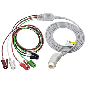
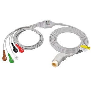
MedLinket's product materials organize veterinary monitoring accessories into dedicated SpO₂, ECG, NIBP, temperature, and EtCO₂ series. The veterinary SpO₂ S-series includes tongue-clip and other site-specific styles. Sensor selection must account for animal anatomy, intended site, connector, cable configuration, and the monitor's oximetry technology—not simply whether the plug appears to fit.
Browse veterinary monitoring supplies. For sensor-style selection, read choosing a multi-site Y-type SpO₂ sensor for animals . For the differences between human and veterinary sensor design, see veterinary vs human SpO₂ sensors .
Compatibility information needed Before ordering, provide the monitor manufacturer and model, oximetry technology, connector photo or part number, intended animal species, preferred site, cable length, and reusable or single-patient-use requirement. Third-party brand names indicate compatibility only, not OEM manufacture, authorization, affiliation, or endorsement.
Why MedLinket emphasizes signal acquisition
- Biomedical signal focus: MedLinket has developed sensors, cables, modules, and monitoring accessories since 2004.
- Defined veterinary accessory families: company product materials separate veterinary SpO₂, ECG, temperature, NIBP, and EtCO₂ accessory series.
- SpO₂ evaluation capability: company materials describe an internally developed FiO₂ simulator, SaO₂ stability evaluation system, and data-acquisition capability. Specific accuracy claims must remain tied to the exact product and report.
- Compatibility labeling: connector and monitor compatibility should be verified for the exact model rather than inferred from a brand name alone.
Learn more on the MedLinket company page and request product-specific technical and compatibility documentation before purchasing.
Frequently asked questions
Why is my veterinary pulse oximeter not reading on a dog or cat?
How do I know whether a veterinary SpO₂ reading is reliable?
Does fur affect a veterinary SpO2 sensor?
Why does a pulse oximeter reading become unstable during anesthesia?
Can pigmentation cause an inaccurate animal SpO₂ reading?
How can I test whether the sensor or cable is faulty?
Can a human finger SpO₂ sensor be used on an animal?
This article provides general educational and product-selection information. It is not a substitute for a veterinarian's examination, diagnosis, treatment, device instructions, or clinical judgment. SpO₂ accuracy varies with the patient, species, perfusion, motion, site, tissue properties, sensor, cable, monitor, algorithm, and calibration range. Reassess the animal whenever a reading is low, absent, unstable, or inconsistent with the clinical condition. Confirm specifications and compatibility for the exact equipment before purchase or use.