By MedLinket Clinical Education Team | Updated: February 2026 | 10 min read
📚 This article is part of our Hospital Monitor Reading & Accessories Guide series — a comprehensive resource covering every parameter you see on a bedside monitor.
Quick Answer: Core temperature measures the thermal state of the body's deep organs and central blood supply (esophageal, rectal, bladder, pulmonary artery, nasopharyngeal). Peripheral temperature is measured at the body surface (axillary, forehead, temporal artery, skin surface). Core temperature is more accurate for clinical decisions. Peripheral readings may be 0.5–2°C lower and are significantly influenced by ambient conditions, perfusion status, and vasoconstriction. As a general rule: use core temperature monitoring for surgical patients, critically ill patients, and anyone at risk for thermoregulatory complications. Use peripheral temperature for general screening and trending in stable ward patients.
Walk into any operating room mid-case and glance at the patient monitor — alongside SpO₂, heart rate, and NIBP, you will see one or two temperature values displayed. For many clinicians, temperature feels like the most straightforward vital sign. But choosing the wrong measurement site — or misinterpreting the reading — can delay intervention for hypothermia, mask a fever in sepsis, or trigger unnecessary treatment. Understanding core vs peripheral temperature is a daily clinical decision that directly impacts patient outcomes.
Core vs Peripheral Temperature: Key Differences at a Glance
A 68-year-old patient post-CABG surgery shows:
Axillary temp: 36.8°C (appears normal)
Esophageal temp: 35.2°C (moderate hypothermia)
Outcome: Delayed extubation, increased bleeding, prolonged PACU stay
Moral: Relying on peripheral temperature in post-op patients can mask dangerous hypothermia.
| Aspect | Core Temperature | Peripheral Temperature |
|---|---|---|
| Normal Range | 36.5–37.5°C (97.7–99.5°F) | Typically 0.5–2°C lower than core |
| Accuracy | High — reflects true internal temperature | Lower — affected by environment and perfusion |
| Response Time | Slower to change (tightly regulated) | Faster to change (environment-dependent) |
| Invasiveness | More invasive (probe insertion required) | Non-invasive or minimally invasive |
| Best Clinical Use | Surgery, ICU, anesthesia, therapeutic hypothermia | General ward screening, outpatient triage |
| Environmental Influence | Minimal | Significant (ambient temp, air flow, clothing) |
| Equipment | Esophageal/rectal/bladder temperature probes | Skin surface temperature sensors, IR thermometers |
Core Temperature Measurement Sites
Core body temperature is tightly regulated by the hypothalamus and normally stays within 36.5–37.5°C. Unlike peripheral readings, core measurements reflect the actual thermal status of blood supplying the heart, brain, and abdominal organs — making them the standard for critical clinical decisions.
Esophageal (Gold Standard for Anesthesia)
The distal esophageal temperature probe sits directly behind the left atrium, tracking true core temperature with minimal lag. The American Society of Anesthesiologists (ASA) recommends temperature monitoring for all patients receiving general anesthesia lasting more than 30 minutes. For accurate readings, the probe tip must be positioned in the lower third of the esophagus — at the point of maximum heart sounds.
✓ Clinical Tip: In intubated patients, esophageal temperature is more reliable than rectal temperature and more practical than tympanic measurement. However, in small children and cachectic patients, high-flow respiratory gases near the tracheobronchial tree can cause erroneous readings if the probe is placed too high. If you encounter unexpected values, follow the same systematic approach used when a patient monitor shows no reading — verify probe placement first, then check the cable and monitor connection.
Rectal
Rectal temperature is commonly used in pediatrics and is considered a reliable core body temperature measurement site at rest. However, it has an important limitation: rectal readings lag behind rapid core temperature changes because the rectum is not as well-perfused as central organs. This means rectal temperature may miss the early stages of malignant hyperthermia or the initial drop during anesthetic induction.
⚠️ Avoid rectal temperature in: immunosuppressed or neutropenic patients (infection risk from mucosal injury), post-rectal surgery, and patients with thrombocytopenia or rectal pathology.
Bladder (Foley Catheter)
Bladder temperature is convenient for catheterized patients. When urine flow is adequate, bladder temperature correlates well with core temperature. However, low urine output causes bladder temperature to lag behind actual core changes — a real concern in dehydrated or hemodynamically unstable patients.
Pulmonary Artery (Most Accurate, Most Invasive)
The pulmonary artery catheter (Swan-Ganz) thermistor provides the most precise core temperature measurement because it directly measures blood from the body's core. However, its invasiveness limits use to cardiac surgery and select critical care patients requiring hemodynamic monitoring via IBP transducers and PA catheters.
Nasopharyngeal
Nasopharyngeal probes are good indicators of cerebral temperature. For accurate readings, the probe should be inserted 10–20 cm in adults. This site carries a small risk of epistaxis, particularly in patients with coagulation abnormalities.
Peripheral Temperature Measurement Sites
Peripheral temperature sensors offer speed and convenience, but they estimate rather than directly measure core temperature. Skin temperature is driven by ambient conditions, local perfusion, and thermoregulatory vasoconstriction — meaning readings can diverge significantly from core values in cold environments, shock, or post-anesthesia redistribution.
Axillary
Axillary measurement is one of the most common non-invasive methods, particularly in general wards. However, axillary temperatures average 0.5–1.9°C lower than core and are highly technique-dependent — the probe must sit directly over the axillary artery with the arm fully adducted.
✓ Pro Tip: If your axillary reading seems inconsistent with the patient's clinical picture (e.g., axillary shows 37.5°C but the patient appears septic), recheck with a more accurate core method. In sepsis recognition, the difference between an axillary 37.5°C and a rectal 38.3°C may determine whether fever criteria are met. A mismatch between temperature and other vital signs — like an unexpectedly elevated heart rate — should prompt reassessment.
Tympanic (Ear)
Tympanic infrared thermometers theoretically reflect core temperature because the tympanic membrane shares blood supply with the hypothalamus. In practice, accuracy depends heavily on technique — improper probe angle, cerumen obstruction, or failing to straighten the ear canal will produce unreliable readings. Avoid tympanic measurement in neonates (ear canals too small) and patients with ear infections or recent ear surgery.
Temporal Artery
Temporal artery scanners are quick and non-invasive — ideal for triage and screening. However, they measure surface temperature of the temporal artery and can be affected by sweat, ambient temperature, and hair. Evidence suggests temporal readings are not reliable enough for clinical decision-making in critical care.
Skin Surface
Skin surface temperature probes (forehead, chest, or extremity) are useful for trending and monitoring core-to-peripheral temperature gradients. Forehead skin temperature can run 2°C or more below core temperature. Skin surface monitoring is valuable for assessing vasoconstriction status and guiding warming therapy in post-operative patients, but should never replace core monitoring in critical situations.
🎯 Core Temperature Required? Quick Check
• General anesthesia >30 min
• Cardiac surgery
• Malignant hyperthermia risk
• Therapeutic hypothermia
• Hemodynamically unstable
• General ward screening • Trending in stable patients
• Routine post-op ward care
• Environmental monitoring
When Core Temperature Monitoring Is Essential
Clinical guidelines are clear about when core monitoring is non-negotiable:
| Clinical Scenario | Recommended Core Site | Why Core Monitoring Matters |
|---|---|---|
| General anesthesia (>30 min) | Esophageal or nasopharyngeal | Anesthetics impair thermoregulation; redistribution hypothermia occurs in first hour |
| Cardiac surgery | Esophageal + rectal/bladder | Deliberate cooling/rewarming requires dual-site monitoring |
| Suspected malignant hyperthermia | Esophageal (fastest response) | Peripheral sites lag and may miss early rapid rise |
| Therapeutic hypothermia (post-cardiac arrest) | Esophageal or bladder | Precise target temperature management (32–36°C) required |
| Trauma patients | Rectal or bladder | Hypothermia worsens coagulopathy and outcomes |
| Neonatal care (NICU) | Rectal or skin with servo-control | Neonates lose heat rapidly; hypothermia increases morbidity |
| Sepsis evaluation | Rectal or oral (electronic) | Peripheral may underestimate fever, delaying sepsis recognition |
Perioperative Hypothermia: Why Temperature Accuracy Is a Patient Safety Issue
Inadvertent perioperative hypothermia (core temperature below 36°C) remains one of the most common and underappreciated surgical complications. Based on clinical feedback from hospitals across 120+ countries that use MedLinket patient monitoring accessories, here is what we consistently see when temperature monitoring is inadequate:
-
Increased surgical site infection risk — even 1–2°C core temperature drop impairs neutrophil function and reduces subcutaneous oxygen tension
-
Coagulopathy — hypothermia inhibits platelet function and the coagulation cascade, increasing blood loss and transfusion requirements
-
Prolonged recovery — patients with perioperative hypothermia have longer PACU stays and delayed anesthetic emergence
-
Cardiovascular events — hypothermia triggers sympathetic activation, increasing the risk of myocardial ischemia
The NICE Clinical Guideline CG65 recommends maintaining perioperative core temperature between 36.5–37.5°C and initiating warming interventions when core temperature falls below 36°C. The key word is core — peripheral readings may show normal values while the patient is already hypothermic centrally. If a temperature alarm sounds on the monitor, always verify the reading with a core site before making clinical decisions. For a broader overview of all hospital monitor alarm meanings, see our dedicated alarm guide.
Temperature Probe Selection: Matching the Probe to the Patient
Selecting the right temperature probe involves matching the measurement site, patient population, and clinical scenario. Here is a practical decision framework:
| Patient Population | Recommended Probe Type | MedLinket Solution |
|---|---|---|
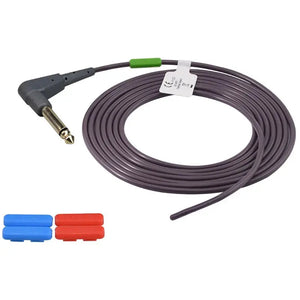
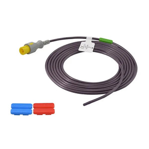
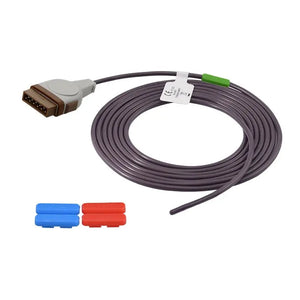
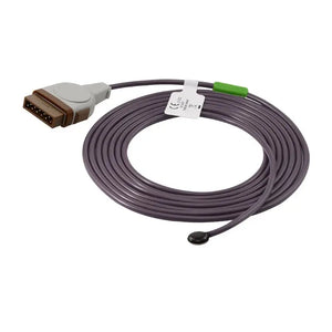
| Adult – General anesthesia | Esophageal or nasopharyngeal probe | Reusable temperature probes (compatible with Philips, GE, Mindray, Dräger) |
| Adult – ICU/ward monitoring | Skin surface or rectal probe | Disposable skin surface probes |
| Neonate – Incubator/warmer | Skin surface probe (servo-controlled) | Dräger-compatible incubator temp probes · Atom-compatible incubator temp probes |
| Neonate – Disposable monitoring | Single-use skin surface probe | Dräger MX11001 disposable temp probes |
Why Probe Compatibility Matters
Temperature probes must match your patient monitor brand's connector type and signal specifications. Using a probe with the wrong impedance characteristics or connector can result in inaccurate readings or a "no reading" alarm — this is one of the most common reasons monitors display "---" for the temperature parameter. For a systematic troubleshooting approach, see our Patient Monitor No Reading Troubleshooting Checklist. The probe's thermistor standard matters too — our bench write-up on how YSI-400 and YSI-700 probes actually behave across different monitor brands shows why a connector that fits is not the whole story.
Not sure which cable or connector matches your monitor? Our cable identification guide walks you through a 5-step method to find the right match. And for the broader comparison of original-equipment accessories vs compatible alternatives, see OEM vs Compatible Accessories: What to Know — and if you're new to non-OEM parts, it helps to be clear on what "compatible" actually means beyond a connector that fits.
MedLinket temperature probes and temperature cables are designed for full signal-level compatibility with major monitoring platforms including Philips, GE Healthcare, Mindray, and Dräger. All probes are manufactured under our ISO 13485-certified quality system and carry CE and FDA clearances. Behind that claim is a defined bench process — see how connector fit, pinout and signal stability are verified before a probe ships.
Three reusable core/skin probes covering the brands and sites discussed above — match to your monitor fleet
Practical Tips: Avoiding Common Temperature Monitoring Mistakes
Based on feedback from clinical engineers and nursing staff — including discussions frequently seen in critical care nursing communities — here are the most common errors and how to prevent them:
Mistake 1: Relying on axillary temperature for critical decisions. Many ICU nurses have shared experiences where axillary readings showed "normal" values while the patient's core temperature (confirmed by esophageal or rectal probe) was well below 36°C. In shock or end-of-life cases, peripheral temperatures can be dramatically lower than core.
Mistake 2: Comparing serial readings from different sites. An axillary reading of 37.2°C cannot be directly compared to a subsequent tympanic reading of 37.8°C. Always use the same site and device for trending. If you must switch methods, document the change.
Mistake 3: Esophageal probe placed too high. If the probe tip is in the upper esophagus rather than the distal third, respiratory gas flow can cool the probe and produce falsely low readings. Position the probe at the point of maximum heart sounds.
Mistake 4: Ignoring the core-peripheral gradient. A widening gradient (>2°C difference between core and skin) suggests vasoconstriction and may indicate hypovolemia, hypothermia, or poor cardiac output — before other hemodynamic parameters change. This is a valuable early warning sign that complements the data from other parameters like NIBP readings and EtCO₂.
Mistake 5: Assuming "no reading" means a broken monitor. When the temperature parameter shows dashes or zeros, the issue is usually the probe or cable — not the monitor itself. Run through the standard checks: is the probe connected? Is the cable intact? Is the probe compatible with this monitor brand? Our guide on monitor display problems covers this in detail, and if the issue is clearly a hardware fault, we have guidance on when to call Biomed vs troubleshoot yourself.
Core-Peripheral Temperature Gradient: A Hidden Clinical Tool
📊 Interpreting the Core-Peripheral Gradient
| Gradient | What It Means | Clinical Action |
| 2–4°C | Normal — adequate perfusion | Continue monitoring |
| >4°C (widened) | Vasoconstriction — hypovolemia, shock, hypothermia | Assess perfusion, consider fluids |
| <2°C (narrowed) | Vasodilation — sepsis, anesthesia, active warming | Evaluate for infection, review meds |
ICU nurse notices:
Core temp: 37.0°C | Skin temp: 32.5°C | Gradient: 4.5°C (widened)
Action: Alerted team to possible developing shock before BP dropped
Result: Early fluid resuscitation, avoided code situation
The core-peripheral gradient often changes before other vital signs.
While most clinicians think of temperature as a single number, the difference between core and peripheral temperature is an important hemodynamic indicator. Under normal thermoregulatory control, core temperature stays between 36.5–37.5°C while peripheral tissue temperature runs 2–4°C lower, depending on ambient conditions and vascular tone.
In critically ill patients, monitoring both core and skin temperature sensors simultaneously provides insight into perfusion status:
-
Normal gradient (2–4°C): Adequate peripheral perfusion
-
Widened gradient (>4°C): Vasoconstriction — consider hypovolemia, hypothermia, or low cardiac output
-
Narrowed gradient (<2°C): Vasodilation — may indicate sepsis, neuraxial anesthesia effect, or active warming
This is why dual-site temperature monitoring (one core + one peripheral) is particularly valuable in ICU and perioperative settings. For continuous skin surface monitoring, the GE-compatible adult skin surface probe ($36, 20% off) provides consistent skin-side readings that pair cleanly with a core esophageal/rectal probe to track the gradient on compatible patient monitor accessories.
Temperature Monitoring for Special Populations
Neonates
Neonates are especially vulnerable to temperature instability. They have a high surface-area-to-body-mass ratio, limited subcutaneous fat, and an immature thermoregulatory system. Unlike adults, neonates cannot shiver and depend on non-shivering thermogenesis (brown fat metabolism) for heat production.
In the NICU, skin temperature probes connected to servo-controlled incubators or radiant warmers are the standard approach. The probe must be securely attached to the skin (typically on the abdomen) to avoid false readings from ambient air. MedLinket's incubator/warmer temperature probes are designed with gentle adhesion and accurate thermistor response for this application. For neonatal SpO₂ monitoring, proper sensor selection is equally critical — learn more in our guide on interpreting ductal sats in neonates.
Pediatric Patients
In pediatrics, rectal temperature remains the most accurate non-invasive core measurement, particularly for infants under 3 months when fever workup decisions are critical. For older children who tolerate it, oral electronic thermometry provides readings within ±0.5°C of core temperature. Tympanic measurement can be used in children over 6 months, but technique is crucial — pulling the pinna down and back (not up and back as in adults) is essential for accurate readings.
Elderly Patients
Older adults (>70 years) often have lower baseline core temperatures and blunted febrile responses. A "normal" temperature of 37.2°C in an elderly patient with suspected infection may actually represent a significant fever relative to their baseline. Always consider the patient's typical temperature when evaluating readings.
Why MedLinket for Temperature Monitoring Accessories?
MedLinket (Shenzhen Med-Link Electronics Tech Co., Ltd) has been manufacturing patient monitoring accessories since 2004. Our temperature product line includes reusable temperature probes, disposable temperature probes, temperature adapter cables, and infant incubator/warmer probes compatible with 30+ monitor brands.
-
FDA 510(k) cleared — 19 product clearances (verify at FDA CDRH database)
-
CE marked (MDR 2017/745) — 48 Class II product categories
-
ISO 13485:2016 certified | NEEQ-listed (Stock Code: 833505)
-
3 owned factories (Shenzhen, Shaoguan, Indonesia) | 3,500+ molds | 16,651+ product variants
-
$5 million USD product liability insurance — individual certificates available for distributors
-
100% factory testing | On-site audits passed from NMPA, FDA, ANVISA, and major OEMs
If you're vetting a supplier rather than just a probe, here's what FDA 510(k), ISO 13485 and the $5M liability figure actually mean in practice — the difference between a certificate on file and verified, audited compliance.
For recommended replacement intervals across all accessory types — temperature probes, SpO₂ sensors, ECG cables, and more — see our Accessory Replacement Schedule.
Frequently Asked Questions
How much lower is peripheral temperature than core?
Peripheral temperature is typically 0.5–2°C lower than core in a stable, normothermic patient. In vasoconstricted states (post-anesthesia, shock, hypothermia), the difference can exceed 4°C. Forehead skin temperature specifically can run 2°C or more below core values. This is why clinical guidelines do not recommend peripheral-only monitoring for patients under anesthesia or in critical care.
Which temperature site is most accurate?
Pulmonary artery temperature is the most accurate core measurement but is too invasive for routine use. For practical purposes, the distal esophageal probe is the gold standard during anesthesia, while rectal temperature is the most reliable non-surgical core site. Among non-invasive methods, only oral and rectal electronic thermometers consistently stay within ±0.5°C of true core temperature. For OR and ICU use, a reusable core probe such as the YSI-compatible esophageal/rectal probe ($36, 40% off) covers both esophageal and rectal placement.
Which site is best for pediatric patients?
For infants under 3 months, rectal temperature is recommended when precise core measurement is needed. For children over 6 months, tympanic measurement is acceptable with proper technique. Axillary is suitable for screening but should be confirmed with a core method if inconsistent with the clinical picture. Always avoid rectal measurement in immunosuppressed children.
Why does my patient's temperature reading seem wrong?
The most common causes are: wrong probe placement, poor skin contact (surface probes), ambient temperature interference (peripheral sites), and probe-monitor incompatibility. Verify probe position, check the cable connection, and confirm probe compatibility. For step-by-step troubleshooting, see our no reading troubleshooting checklist.
How often should temperature probes be replaced?
Disposable temperature probes are single-use. Reusable probes should be inspected before each use for cable damage, connector corrosion, and probe tip integrity. Most reusable temperature probes have a typical service life of 12–18 months with proper handling. Replace immediately if readings become erratic. See our accessory replacement schedule for detailed guidance by parameter type.
Related Articles in This Series
This article is part of the Hospital Monitor Reading & Accessories Guide. Explore related topics:
Medical Disclaimer: This article is intended for educational purposes for healthcare professionals and clinical staff. It does not constitute medical advice. Always follow your facility's protocols and consult qualified medical professionals for patient care decisions.
Need compatible temperature probes for your facility? Contact MedLinket for free compatibility verification and pricing: shopify@medlinket.com | WhatsApp: +852 6467 3105


