By MedLinket Clinical Education Team | Updated: February 2026 | 12 min read
📚 This article is part of our Hospital Monitor Reading & Accessories Guide series. For the complete overview of all alarm types, see Hospital Monitor Alarms: What Each Alarm Means.
⚡ Quick Answer: When the SpO₂ low alarm sounds:
(1) Look at the patient — are they in visible respiratory distress?
(2) Check the pleth waveform — a clean, regular waveform with a consistently low reading suggests true hypoxemia; a poor or absent waveform suggests a sensor problem.
(3) If the patient is truly hypoxic, administer supplemental oxygen per protocol and call for help. Many SpO₂ low alarms are false, caused by poor sensor contact, patient movement, cold extremities, or an aging SpO₂ sensor — but every alarm must be treated as real until verified.
🔑 Key Takeaways
-
The plethysmographic (pleth) waveform is your best tool for differentiating true hypoxemia from a false SpO₂ alarm.
-
Always assess the patient before the equipment: look for cyanosis, dyspnea, altered mental status, and increased work of breathing.
-
The six most common false alarm causes: poor sensor placement, patient movement, cold extremities, nail polish, ambient light, and degraded sensors.
-
True causes include respiratory depression (especially opioid-related), pneumonia, COPD exacerbation, pulmonary embolism, and airway obstruction.
-
Quality pulse oximeter sensors with proper technology matching dramatically reduce false alarms — sensor-monitor compatibility goes beyond connector fit.
-
SpO₂ <90% in a symptomatic patient is a clinical emergency requiring immediate intervention.
Step-by-Step Response: What to Do When SpO₂ Drops
📋 Case in point: Post-op patient, SpO₂ alarm reading 89%. The nurse runs the three-step protocol:
- Patient alert, breathing comfortably, no cyanosis
- Pleth waveform erratic with spikes (motion artifact)
- Patient shivering continuously, ice-cold fingers
→ Switched to ear clip sensor. SpO₂ immediately reads 96%, alarm resolved.
Speed matters when the SpO₂ alarm sounds — but systematic speed matters more than panicked speed. This three-step algorithm applies whether you're in the ICU, a med-surg floor, the PACU, or during transport.
🔔 SpO₂ LOW ALARM SOUNDS
↓
STEP 1: LOOK AT THE PATIENT (3-second visual assessment)
→ Visible cyanosis? (lips, fingertips, nail beds)
→ Dyspneic? (labored breathing, nasal flaring, accessory muscles)
→ Responsive and oriented?
↓
SIGNS OF DISTRESS → Apply/increase supplemental O₂, elevate HOB, call for help, stay with patient
NO DISTRESS → Proceed to Step 2
↓
STEP 2: CHECK THE PLETH WAVEFORM
→ Clean, regular, well-formed waveform? Pulse rate matches ECG heart rate?
↓
GOOD WAVEFORM + LOW READING → TRUE LOW SpO₂. Clinical intervention needed. Assess airway, breathing, circulation. Notify physician.
POOR/ABSENT WAVEFORM → Proceed to Step 3
↓
STEP 3: CHECK THE SENSOR
→ SpO₂ sensor properly placed? Finger warm with adequate perfusion? Nail polish? Sensor intact? Cable connected?
↓
Fix the issue → Reassess in 15–30 seconds
Still low + poor waveform → Try alternate site (ear, toe, different finger)
Still low + good waveform → TREAT AS TRUE HYPOXEMIA
⚠️ Critical Rule: A patient can be alert, conversational, and appear comfortable with an SpO₂ of 85%. Some patients — particularly those with chronic hypoxemia — compensate remarkably well. Always verify with the pleth waveform. If the waveform is good and the reading is consistently low, the patient is desaturating regardless of appearance.
True Hypoxemia vs False Alarm: The Pleth Waveform Is Your Answer
The plethysmographic (pleth) waveform — the pulsating wave alongside the SpO₂ number — shows whether the pulse oximeter is getting a clean, reliable arterial signal. A normal pleth waveform has a sharp steep upstroke (systole), a dicrotic notch (aortic valve closure), and a smooth downslope (diastole). When this pattern is clean, the SpO₂ number is reliable.
🔴 Signs of True Hypoxemia
-
Good pleth waveform with consistent low reading
-
Visible cyanosis (blue/dusky lips, fingertips)
-
Increased work of breathing — tachypnea, nasal flaring, accessory muscles
-
Altered mental status — restlessness, confusion, agitation
-
Tachycardia (compensatory heart rate increase)
-
Reading does not change when sensor is repositioned
🔵 Signs of a False Alarm (Sensor Problem)
-
Poor, erratic, or absent pleth waveform
-
Patient appears comfortable, well-perfused, no distress
-
SpO₂ jumps erratically (e.g., 99% → 72% → 95%)
-
Sensor feels loose or has shifted position
-
Cold or edematous extremity at sensor site
-
HR on SpO₂ doesn't match HR on ECG
-
Reading improves when sensor is moved to another site
💡 Clinical Pearl — The HR Match Test: Compare the pulse rate from the SpO₂ sensor with the heart rate from the ECG. If they match closely, the sensor is detecting a valid arterial pulse and the SpO₂ reading is likely reliable. If they diverge significantly, the sensor is struggling. This 2-second check saves enormous diagnostic uncertainty.
🧠 Quick troubleshooting mnemonic — "MOVE PLACE":
- Movement → Have patient hold still, consider ear clip
- Orientation → Check LED-detector alignment
- Vascular → Check perfusion index, warm the extremity
- Equipment → Swap in a new sensor to test
- Position → Verify fingertip reaches sensor end
- Light → Block ambient light interference
- Acrylic → Remove nail polish/gel
- Circulation → Improve peripheral perfusion
- Expired → Check sensor expiration/condition
6 Common Causes of False Low SpO₂ Alarms
Pulse oximetry relies on light transmission through pulsating arterial blood. Anything that disrupts the light path, dampens the pulse signal, or introduces optical noise degrades the reading.
1. Poor Sensor Placement (Most Common)
The LED emitter must be directly opposite the photodetector with the tissue centered between them. When the sensor shifts, light bypasses tissue — optical shunting — producing inaccurate readings. For disposable SpO₂ sensors, verify the adhesive wrap isn't too loose or overlapping the optical window.
Fix: Reposition the sensor. Ensure fingertip reaches the end. For pediatric patients, try the toe. For neonates, a foot-wrap sensor provides better contact than a finger clip.
2. Patient Movement (Motion Artifact)
Motion causes venous blood to slosh, creating pulsatile venous flow the oximeter misinterprets as arterial pulsation. Since venous blood has lower oxygen saturation, this produces a falsely low reading. Especially prevalent during transport, in the PACU, and with pediatric patients.
Fix: Wait for patient to settle. For chronic restlessness, consider an ear clip sensor — ear measurement is less affected by extremity motion. Advanced SpO₂ sensors with motion-resistant algorithms (Masimo SET, Nellcor OxiMax) filter motion artifact.
3. Cold Extremities / Poor Peripheral Perfusion
Pulse oximetry depends on detecting pulsatile arterial blood flow. When peripheral perfusion is poor — due to hypothermia, vasoconstriction, vasopressors, or peripheral vascular disease — the pulse amplitude is too weak for reliable measurement. The Perfusion Index (PI) on many monitors quantifies pulse signal strength — a PI below 0.3 generally indicates insufficient perfusion. For context on how vasoconstriction affects monitoring, see our guide on core vs peripheral temperature and the core-peripheral gradient.
Fix: Warm the patient's hand. Try the ear lobe (centrally perfused). For patients on high-dose vasopressors, a forehead reflectance sensor may be necessary.
4. Nail Polish, Acrylic Nails, and Gel Nails
Dark nail polish (especially blue, black, dark green) absorbs 660nm red light, mimicking deoxygenated hemoglobin absorption and producing falsely low SpO₂.
Fix: Remove nail polish from the measurement finger, or rotate the sensor 90° so light passes through the sides of the finger. Alternatively, use an adjacent finger, the ear, or a toe.
5. Ambient Light Interference
Strong ambient light — surgical lights, fluorescent fixtures, warming lamps (neonatal units), or direct sunlight — floods the photodetector and corrupts the signal.
Fix: Cover the sensor with an opaque material. Reposition the hand away from direct light.
6. Degraded or Incompatible Sensor
Over time, LEDs in reusable SpO₂ sensors degrade, housings crack, and cable shielding breaks down. Equally critical: a sensor whose connector physically fits but uses a different pulse oximetry technology will produce unreliable readings. A Nellcor OxiMax sensor will not give accurate readings on a Masimo SET module, even if the connector mates.
Fix: Replace damaged or expired sensors (12–18 month typical lifespan). Confirm sensor technology matches the monitor's SpO₂ module. See our OEM vs compatible accessories guide and cable identification guide for matching methods.
💡 From MedLinket's Compatibility Lab: We encounter "connector fits but technology doesn't match" more often than expected. One hospital found SpO₂ readings consistently 3–5% lower than ABG values with doubled false alarms overnight. Root cause: the sensors used a different signal protocol than the monitor expected. After switching to properly matched compatible SpO₂ sensors, alarm rates returned to baseline immediately.
Clinical Causes of True Low SpO₂
When you've confirmed a good pleth waveform and a consistently low reading, the patient is genuinely desaturating:
| Clinical Cause | Typical Presentation | Key Assessment | Initial Action |
|---|---|---|---|
| Respiratory depression (opioid/sedative) | Gradual SpO₂ decline, often post-surgery | RR <8/min, shallow breathing, somnolence, pinpoint pupils | Stimulate patient, O₂, call physician; have naloxone available |
| Pneumonia | Progressive desaturation over hours to days | Fever, productive cough, crackles, increased RR | Apply O₂, position upright, monitor trend, notify physician |
| COPD exacerbation | SpO₂ chronically lower at baseline (88–92%); acute drop | Wheezing, prolonged expiration, accessory muscles | Controlled low-flow O₂ (target 88–92%), bronchodilators |
| Pulmonary embolism | Sudden acute desaturation | Sudden dyspnea, pleuritic chest pain, tachycardia, anxiety | High-flow O₂, keep patient still, emergent notification |
| Heart failure / pulmonary edema | Worsens supine, improves sitting | Bilateral crackles, pink frothy sputum, JVD, edema | Position upright, O₂, restrict fluids, emergent notification |
| Airway obstruction | Sudden desaturation, may be rapid | Stridor, absent breath sounds, inability to speak | Open airway, suction if mucus, call code if complete obstruction |
| Atelectasis | Post-operative, common Day 1–3 | Diminished breath sounds, shallow breathing, mild fever | Deep breathing / incentive spirometry, reposition, O₂ |
| Sleep apnea (OSA) | Intermittent nocturnal desaturation | Snoring, apneic episodes, SpO₂ dips then recovers | Lateral position, consider CPAP, document pattern |
⚠️ Opioid-Induced Respiratory Depression: The pattern is insidious — patient receives pain medication, becomes drowsy, RR drops, SpO₂ slowly declines over 15–30 minutes. If the alarm is attributed to "sensor issue," the intervention window narrows dangerously. When a post-surgical patient's SpO₂ alarms, always check respiratory rate and consciousness before the sensor.
If the patient also shows a widening core-peripheral temperature gradient or falling blood pressure, consider a more systemic cause like sepsis or hemorrhage rather than isolated respiratory pathology.
Special Populations: Adjusted SpO₂ Targets
Default alarm thresholds cause constant false alarms for some populations and insufficient warning for others:
| Patient Population | Normal SpO₂ Range | Suggested Low Alarm | Clinical Rationale |
|---|---|---|---|
| Healthy adult | 95–100% | 90% | Standard threshold |
| COPD patient | 88–92% | 85–88% (per physician) | Excessive O₂ may suppress respiratory drive |
| Premature neonate (on O₂) | 91–95% target | 88% low / 95% high | Hyperoxemia risks retinopathy of prematurity |
| Full-term newborn (first 10 min) | 60–90% (rising) | Per neonatal protocol | SpO₂ rises progressively after birth |
| Post-surgical patient on opioids | 95–100% | 92% (earlier warning) | Higher threshold detects respiratory depression sooner |
| High altitude | 90–95% | 88% | Lower ambient O₂ reduces baseline |
Neonatal targets carry the tightest tolerances of any population: too little oxygen risks hypoxic injury, while too much risks retinopathy of prematurity. That makes a correctly-sized, well-aligned neonatal sensor — one that conforms to a tiny digit and holds optical alignment despite movement — essential to keeping readings inside the narrow target window.

When to Escalate: Red Flags
🚨 Escalate Immediately If:
-
SpO₂ remains <90% despite confirming good pleth waveform
-
SpO₂ remains <88% on supplemental oxygen
-
Patient shows visible respiratory distress: cyanosis, severe dyspnea, stridor, apnea
-
Respiratory rate <8 or >30 breaths/minute
-
Patient is unresponsive or has altered mental status
-
SpO₂ is dropping rapidly (95% → 85% within minutes)
-
Patient is post-operative and somnolent with recent opioid administration
-
You cannot identify the cause after basic troubleshooting
When escalating, use SBAR: "This is [name], caring for [patient] in [room]. Their SpO₂ has dropped to 84% on room air with a good pleth waveform. They have a history of COPD and were admitted for pneumonia. They are tachypneic at 28/min and mildly confused. I've elevated HOB and applied 2L NC. I'm requesting a physician assessment."
Preventing Recurring SpO₂ False Alarms
If a patient's sensor alarms repeatedly and you've confirmed it's not true hypoxemia, solve the problem systematically:
-
Verify sensor-monitor compatibility. Physical connector fit does not guarantee signal compatibility. Check with your biomedical department or the sensor compatibility guide.
-
Choose the right sensor type. Standard finger clips for alert, warm adults. Wrap sensors for neonates. Ear clips for poor perfusion. Adhesive sensors for restless patients. See: Understanding SpO₂ Sensors: Masimo, Nellcor & Neonatal Options.
-
Optimize sensor site. Rotate between fingers every 2 hours. Avoid fingers with nail polish, poor perfusion, or edema. Index or middle finger typically provides the best signal.
-
Customize alarm thresholds. A COPD patient with baseline SpO₂ of 89% should not alarm at 90%. Research shows lowering the threshold from 90% to 88% with a 15-second delay can reduce SpO₂ alarms by over 80% without compromising safety.
-
Use alarm delays appropriately. 10–15 second delays filter transient events (sleep, repositioning). Collaborate with your unit's alarm committee.
-
Replace degraded sensors. Reusable SpO₂ sensors have a 12–18 month lifespan. Inspect regularly for cracked housings, discolored LEDs, and loose connectors.
For the most common scenario — routine continuous monitoring of an alert, warm adult — a comfortable reusable soft-silicone finger sensor is the everyday workhorse. It seats more securely and stays gentler over long wear times than a rigid clip, which means fewer movement-driven dropouts and fewer false alarms over a shift.
🔗 MedLinket SpO₂ Sensors: Reducing False Alarms at the Source
MedLinket's SpO₂ sensors achieve ±2% accuracy across 70–100% SpO₂ and are individually validated against the specific monitor module — not just the connector.
-
Patented over-temperature protection — automatically disengages at skin temperatures above 41°C, preventing burns (especially critical for neonates)
-
Multi-adapter strategy: One sensor probe + multiple adapter cables = compatibility with Mindray, Philips, GE, Masimo, Nellcor, and more — reducing SKU complexity by up to 60%
-
Full regulatory compliance: FDA 510(k), CE MDR, ISO 13485:2016
Product links:
-
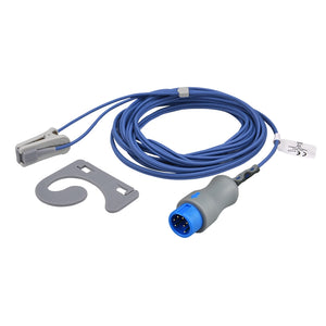
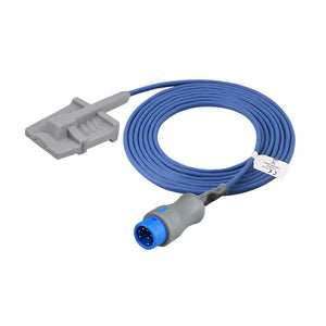
Ear Clip SpO₂ Sensor (for poor perfusion / motion) → ($56.00, 20% off)
-
Adult Soft Finger SpO₂ Sensor (standard monitoring) → ($56.00, 20% off)
-
Neonatal Silicone Wrap SpO₂ Sensor → ($56.00, 20% off)
Why Hospitals Trust MedLinket
MedLinket (Shenzhen Med-Link Electronics Tech Co., Ltd., NEEQ: 833505) has manufactured patient monitor accessories since 2004. Our SpO₂ sensors, monitoring accessories, and disposable SpO₂ sensors are used in 2,000+ hospitals across 120+ countries.
-
FDA 510(k) cleared (19 clearances) | CE MDR | ISO 13485:2016
-
3 factories (Shenzhen, Shaoguan, Indonesia) | 3,500+ molds | 16,651+ product variants
-
$5M USD product liability insurance — individual certificates available
-
45 utility model patents + 8 invention patents including over-temperature protection
Need compatibility verification? Send us your monitor model: shopify@medlinket.com | WhatsApp: +852 6467 3105
Frequently Asked Questions
At what SpO₂ level should I be concerned?
For most patients, SpO₂ below 94% warrants attention, below 90% requires active intervention, and below 85% is a clinical emergency. However, the trend matters as much as the number — a patient dropping from 97% to 93% over 30 minutes may be more concerning than a COPD patient stable at 89%. Always assess in context.
Why does the SpO₂ alarm go off more at night?
Several factors converge: physiologic desaturation during sleep (healthy adults may dip to 93–94% in deep sleep), obstructive sleep apnea causing episodic desaturations, opioid effects peaking during nighttime dosing, and patients shifting position causing sensor displacement. Consider slight threshold adjustments (per physician order) and adhesive sensors that stay in place.
How do I know if a low SpO₂ reading is real or a sensor problem?
Check the pleth waveform. A clean, regular waveform with a consistent low SpO₂ strongly indicates true hypoxemia. A poor, erratic, or absent waveform indicates a sensor or perfusion problem. Also check whether the pulse rate on SpO₂ matches the heart rate on ECG — agreement suggests a valid signal. If uncertain, check ABG.
Can SpO₂ be falsely high?
Yes. The most significant cause is carbon monoxide poisoning. Carboxyhemoglobin absorbs 660nm red light almost identically to oxyhemoglobin, so a standard pulse oximeter reads CO-bound hemoglobin as oxygenated. A patient with severe CO poisoning can show SpO₂ of 98–100% while oxygen-carrying capacity is critically compromised. In suspected CO exposure, obtain an ABG with co-oximetry.
Does dark skin pigmentation affect SpO₂ accuracy?
Yes. Published research shows standard pulse oximeters can overestimate SpO₂ by 2–3% in patients with darker skin pigmentation, particularly at lower saturation levels. A patient displaying SpO₂ of 92% may have true saturation closer to 89–90%. Be vigilant for clinical signs of hypoxemia even when the number appears borderline-acceptable.
What SpO₂ sensor type is best for neonates?
Wrap-style (silicone wrap) sensors provide the most reliable neonatal readings because they conform to small digits and maintain optical alignment despite movement. In the delivery room, right hand (pre-ductal) placement is standard. MedLinket's neonatal sensors include patented over-temperature protection that disengages at 41°C. For more on neonatal SpO₂, see Interpreting Ductal Sats in Neonates.
How often should I change the SpO₂ sensor site?
Rotate every 2 hours during continuous monitoring to prevent pressure injury. Inspect the previous site for irritation. Choose a well-perfused digit without edema or nail polish. For full replacement intervals across all accessory types, see the Accessory Replacement Schedule.
Related Articles in This Series
This article is part of the Hospital Monitor Reading & Accessories Guide. Explore related topics:
Medical Disclaimer: This article is intended for educational purposes for healthcare professionals and clinical staff. It does not constitute medical advice. Always follow your facility's protocols and consult qualified medical professionals for patient care decisions.
Declaration: All product names, brands, and OEM part numbers (e.g., Mindray, Philips, GE, Masimo, Nellcor, Edan, and associated model numbers) are used for identification and compatibility-reference purposes only and do not imply any affiliation, partnership, or endorsement. MedLinket products are independently verified compatible alternatives. Product images and actual objects may differ slightly in appearance (e.g., connector design, color) but function the same.


