Heart rate alarms on a hospital monitor indicate rates outside set limits. A high HR alarm (tachycardia, >100 BPM in adults) may signal pain, fever, anxiety, dehydration, or cardiac arrhythmia. A low HR alarm (bradycardia, <60 BPM) may be normal in athletes and during sleep, or may indicate heart block, medication effects, or vagal response. Always assess the patient first — then check the equipment.
Heart rate alarms are among the most frequent alerts on patient monitors in ICU, telemetry, and general medical-surgical units. Research from the MIMIC II ICU database found that extreme tachycardia and bradycardia alarms together accounted for nearly 48% of all critical ECG arrhythmia alarms — yet a significant portion were false positives triggered by artifact. Knowing how to quickly distinguish a real alarm from a false one is essential for every bedside clinician.
Heart Rate Alarm Response: Always Start With the Patient
Before touching any button on the monitor, look at your patient. This prevents two dangerous errors: ignoring a real emergency and wasting time troubleshooting equipment while a patient deteriorates.
| Step | Action | If Yes | If No |
|---|---|---|---|
| 1. Look at the patient | Signs of distress? (altered consciousness, diaphoresis, chest pain, pallor) | → Stay with patient, call for help, begin intervention | → Proceed to Step 2 |
| 2. Check the ECG waveform | Clean waveform with identifiable P-QRS-T morphology? | → Likely true alarm; assess clinically, notify provider | → Likely artifact; proceed to Step 3 |
| 3. Compare HR vs PR | Does HR from ECG match pulse rate from SpO₂ sensor? | → Consistent readings support a true alarm | → Discrepancy suggests electrode or sensor issue |
| 4. Check equipment | Are ECG electrodes and ECG lead wires securely attached? | → Equipment OK; re-assess clinically | → Reattach or replace; re-assess after fix |
Cross-reference the heart rate from ECG (labeled "HR") against the pulse rate from the pulse oximeter (labeled "PR"). When both match, it strongly supports a true reading. When they diverge — especially if the SpO₂ waveform looks clean but the ECG trace is noisy — the issue is almost always on the ECG side.
High Heart Rate (Tachycardia) Alarm: Common Causes
When heart rate exceeds 100 BPM, the monitor triggers a tachycardia alarm. Most monitors use a default high limit of 120–130 BPM (warning) and 140 BPM (critical/red).
| Cause | Associated Signs | Typical Intervention |
|---|---|---|
| Pain | Patient report, grimacing, guarding, elevated BP | Pain assessment, analgesics per order |
| Fever / infection | Elevated temperature, diaphoresis, elevated WBC | Cultures, antipyretics, antibiotics as ordered |
| Anxiety / agitation | Restlessness, rapid respirations | Reassurance, anxiolytics if indicated |
| Hypovolemia / dehydration | Dry mucous membranes, low urine output, orthostatic changes, low blood pressure | IV fluid bolus, intake/output assessment |
| Medication effect | Recent administration of stimulant, beta-agonist, or vasoactive drug | Review medication timing, notify provider |
| Cardiac arrhythmia (SVT, AFib, VTach) | Irregular rhythm on ECG, palpitations, hemodynamic instability | 12-lead ECG, ACLS protocol, urgent provider notification |
When High HR Is Dangerous — Escalate Immediately
-
Sustained HR >150 BPM with hemodynamic compromise (hypotension, altered mental status)
-
Wide QRS complex (>0.12 seconds), suggesting ventricular tachycardia
-
Patient reports chest pain, syncope, or severe dyspnea
-
Irregularly irregular rhythm at a rapid rate (atrial fibrillation with rapid ventricular response)
Ventricular tachycardia (VTach) deserves special attention. Published research found VTach accounted for approximately 80% of critical arrhythmia alarms in ICU — yet 80–90% were false positives, making it a major contributor to alarm fatigue. A clean ECG waveform with identifiable wide QRS morphology is key to confirming true VTach versus artifact.
Low Heart Rate (Bradycardia) Alarm: Common Causes
Most monitors trigger a bradycardia warning at <50 BPM and a critical alarm at <40 BPM. A low heart rate is not always pathological.
| Cause | Associated Signs | Clinical Significance |
|---|---|---|
| Athletic conditioning | Patient otherwise healthy, asymptomatic | Normal variant; HR 40–60 BPM at rest |
| Sleep | Patient sleeping, normal respiratory pattern | Physiological; HR can drop into the 40s during deep sleep |
| Beta-blocker / calcium channel blocker | Known medication history, stable BP | Expected pharmacological effect |
| Vagal response | During vomiting, bearing down, suctioning | Typically transient; monitor closely |
| Heart block (1st, 2nd, 3rd degree) | Irregular or dissociated P-QRS relationship | May require atropine, pacing; notify provider |
| Acute MI (especially inferior wall) | ST changes in leads II, III, aVF; chest pain | Activate STEMI protocol if indicated |
When Low HR Is Dangerous — Escalate Immediately
-
HR <40 BPM with symptoms — dizziness, syncope, altered mental status, diaphoresis
-
Accompanied by hypotension (systolic <90 mmHg or MAP <65 mmHg)
-
Signs of poor perfusion (cool/mottled extremities, capillary refill >3 seconds)
-
New heart block pattern on ECG waveform
-
Abrupt drop (e.g., HR 90 → 51) rather than chronic resting bradycardia
@thecardiologytutor Replying to @Sa Many people worry when they see a low HR. But do you need to worry??? Let me explain… #bradycardia #arrhythmia #cardiology #doctor #cardiologist #ecg #residentdoctor #medstudent #nursingstudent ♬ original sound - TheCardiologyTutor
False Heart Rate Alarms: Causes and Solutions
For heart rate alarms specifically, MIMIC II data found false alarm rates of approximately 29% for extreme bradycardia and 23% for extreme tachycardia. While these are among the more accurate alarm types, they still contribute substantially to overall alarm fatigue.
| Cause of False HR Alarm | Why It Happens | Solution |
|---|---|---|
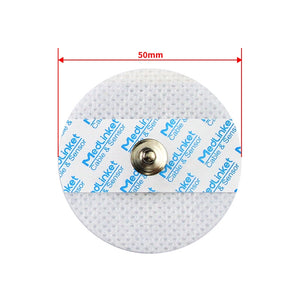
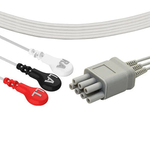
| Loose or dried-out ECG electrodes | Impedance increases → monitor under-counts QRS (false bradycardia) or detects artifact as beats (false tachycardia) | Replace electrodes every 24 hours; use quality disposable ECG electrodes |
| Patient movement / muscle artifact | High-frequency EMG noise mimics QRS, causing over-counting | Secure ECG lead wires; see ECG leads off alarm fix |
| Low-voltage QRS | Monitor cannot detect small QRS → false bradycardia/asystole | Reposition chest electrode to lead with highest QRS voltage |
| T-wave double-counting | Tall T-waves misinterpreted as QRS → HR displayed ~double true rate | Adjust QRS detection sensitivity; select lead with smaller T-waves |
| Electrical interference (50/60 Hz) | Nearby electrical devices disrupt rhythm detection | Move devices away; verify grounding |
| Damaged or incompatible ECG cables | Broken shielding or poor connector contact → intermittent signal dropout | Inspect cables; replace with compatible ECG cables |
Why Equipment Quality Matters
A 2019 FDA report identified cardiac monitor alarms as the leading cause of alarm-related patient deaths. Every false alarm contributes to alarm fatigue. The quality of your monitoring accessories — electrodes, ECG lead wires, and ECG trunk cables — directly affects signal fidelity and false alarm rates.
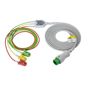
MedLinket's in-house testing shows ECG electrode AC impedance as low as 109Ω — well below the ≤2kΩ industry standard (YY/T 0196-2005) — translating directly into cleaner signals and fewer artifact-triggered alarms. Our offset (eccentric) ECG electrodes reduce motion artifact from cable tension with a patented neck-relief structure, making them ideal for telemetry and Holter monitoring where patients are ambulatory. On the cable side, intermittent dropout often traces back to a marginal connector — see how connector fit and signal stability are verified before a cable ships, since a loose or high-impedance contact is a frequent hidden source of false brady/tachy alarms.
🔗 Products That Reduce False Heart Rate Alarms
-
Disposable ECG electrodes — Offset design reduces motion artifact
-
Mindray compatible 3-lead ECG cable — Shielded construction minimizes EMI
-
Philips compatible ECG leadwires — Secure snap/grabber connections
-
GE compatible ECG leadwires — Molded design for durability
Not sure whether to choose OEM or compatible accessories? See our guide on OEM vs Compatible Patient Monitor Accessories. Need help matching cables to your monitor brand? Use the 5-step cable identification method. And if you're new to non-OEM parts, it's worth being clear on what "compatible" actually means beyond a connector that fits.
Heart Rate Alarm Limits: Default vs Patient-Specific
Published research from a quaternary care ICU found that 50% of patients had alarm settings left at default for their entire stay — even when their baseline fell outside those limits.
| Patient Scenario | Suggested Low | Suggested High | Rationale |
|---|---|---|---|
| General adult | 50 BPM | 120 BPM | Standard default |
| Patient on beta-blocker | 40–45 BPM | 120 BPM | Avoids nuisance alarms from expected bradycardia |
| Young, athletic patient | 40 BPM | 120 BPM | Athletes commonly have resting HR in 40s–50s |
| Post-surgical (early recovery) | 50 BPM | 110 BPM | Tighter high limit to catch early bleeding/hypovolemia |
| Chronic atrial fibrillation | 50 BPM | 130–140 BPM | Wider high limit for expected rate variability |
| Newborn / infant | 100 BPM | 180 BPM | Neonatal normal: 120–160 BPM |
Always follow your facility's policy on alarm limit adjustments. Document every change in the patient's record.
Sinus Rhythm vs Arrhythmia: Quick ECG Guide
When a heart rate alarm fires, glancing at the ECG waveform helps quickly determine if the rhythm is sinus or an arrhythmia requiring urgent attention:
| Rhythm | Rate | Key ECG Features | Urgency |
|---|---|---|---|
| Sinus tachycardia | 100–150 BPM | Normal P-QRS-T, regular, gradual onset | Low–Moderate |
| SVT | 150–250 BPM | Narrow QRS, P-waves hidden, abrupt onset | Moderate–High |
| Atrial fibrillation (rapid) | 100–180 BPM | Irregularly irregular, no identifiable P-waves | Moderate |
| Ventricular tachycardia | >100 BPM | Wide QRS (>0.12s), often regular | High–Critical |
| Sinus bradycardia | <60 BPM | Normal P-QRS-T, regular | Low if asymptomatic |
| 2nd degree block (Mobitz II) | Variable | Some P-waves without QRS; no PR prolongation | High |
| 3rd degree (complete) block | Usually <45 BPM | P-waves and QRS at independent rates | Critical |
Normal Heart Rate Ranges by Age
One of the most common causes of unnecessary alarm escalation is unfamiliarity with age-specific norms. A heart rate of 140 BPM in an adult is tachycardia; in a 6-month-old, it is perfectly normal.
| Age Group | Normal Resting HR | Bradycardia Threshold | Tachycardia Threshold |
|---|---|---|---|
| Newborn (0–28 days) | 120–160 BPM | <100 BPM | >180 BPM |
| Infant (1–12 months) | 100–160 BPM | <100 BPM | >170 BPM |
| Toddler (1–3 years) | 90–150 BPM | <80 BPM | >150 BPM |
| Child (4–12 years) | 70–120 BPM | <70 BPM | >130 BPM |
| Adolescent / Adult | 60–100 BPM | <60 BPM | >100 BPM |
| Trained athlete | 40–60 BPM | Context-dependent | >100 BPM |
Neonatal monitoring requires particular care with alarm limits. Neonatal SpO₂ monitoring considerations apply alongside heart rate — the two should always be interpreted together in the NICU setting. For temperature monitoring, neonates also require special probe selection and tighter thresholds.
Why Hospitals Trust MedLinket ECG Accessories
MedLinket (Shenzhen Med-Link Electronics Tech Co., Ltd., NEEQ: 833505) has manufactured patient monitor accessories since 2004. Our ECG cables, ECG lead wires, and ECG electrodes are used in 2,000+ hospitals across 120+ countries.
-
FDA 510(k) cleared (19 clearances) | CE MDR | ISO 13485:2016
-
ECG electrode AC impedance as low as 109Ω — well below ≤2kΩ standard
-
Offset electrode patent — absorbs cable tension to prevent motion artifact
-
30+ compatible brands: Philips, GE, Mindray, Dräger, Nihon Kohden, and more
-
$5M USD product liability insurance — individual certificates available
If you're vetting a supplier rather than just a part, here's what FDA 510(k), ISO 13485 and the $5M liability figure actually mean in practice.
For recommended cable and electrode replacement intervals, see our Accessory Replacement Schedule. When the issue goes beyond accessories, our guide on When to Call Biomed vs Troubleshoot Yourself helps clinical staff decide when engineering support is needed.
📩 Contact: shopify@medlinket.com | WhatsApp: +852 6467 3105
Frequently Asked Questions
Is it normal for heart rate to change with breathing?
Yes. Sinus arrhythmia — a slight heart rate increase during inspiration and decrease during expiration — is a normal physiological variation, especially common in younger patients. It does not require treatment.
Why does the heart rate alarm go off during sleep?
Heart rate naturally decreases during sleep due to increased parasympathetic tone. Sleeping HRs in the 40s–50s are common. If the monitor's low alarm is set at 50 BPM, it will trigger repeatedly overnight. Discuss adjusting the low limit for patients with known, asymptomatic nocturnal bradycardia.
What if HR and PR show different numbers?
HR (from ECG leads) and PR (from SpO₂ sensor) should match within a few BPM. A significant discrepancy usually means one source has poor signal quality. In patients with PVCs, some beats may not generate a strong enough pulse for the oximeter, so PR will read lower than HR.
Is 55 BPM dangerous?
Not necessarily. If the patient is asymptomatic — alert, oriented, stable blood pressure, warm extremities — 55 BPM is likely safe. However, if symptomatic (dizzy, confused, hypotensive), it warrants clinical assessment and provider notification.
How can our facility reduce heart rate false alarms?
Implement a multi-pronged approach: (1) use quality ECG cables and electrodes with low impedance — for example, our adult 4.0 mm snap ECG electrodes ($11.90/bag of 50, 21% off) tested to ~109 Ω, (2) individualize alarm limits per patient, (3) change electrodes every 24 hours, (4) ensure proper skin preparation, and (5) provide ongoing staff education. For a broader perspective, see our guide on false alarm prevention strategies.
Medical Disclaimer: This article is intended for educational purposes for healthcare professionals and clinical staff. It does not constitute medical advice. Always follow your facility's protocols and consult qualified medical professionals for patient care decisions.


