⚡ Quick answer
SpO2 (peripheral capillary oxygen saturation) measures the percentage of oxygen-carrying hemoglobin in your blood. A normal SpO2 level is 95–100% for healthy adults. Readings below 90% are generally considered low and may need prompt medical attention. SpO2 is displayed on a hospital patient monitor and measured non-invasively by a pulse oximeter — a small sensor on the finger, toe, or earlobe that uses two wavelengths of light (660 nm red and 940 nm infrared) to calculate oxygen saturation in real time.
📋 Key takeaways
- SpO2 stands for peripheral capillary oxygen saturation — the percentage of hemoglobin molecules carrying oxygen in arterial blood.
- Normal SpO2 is 95–100% for healthy adults. COPD patients may have an individualized target around 88–92% per physician orders.
- SpO2 below 90% is a medical concern; below 85% is a medical emergency requiring immediate intervention.
- Regular SpO2 monitoring enables early detection of respiratory deterioration and supports earlier intervention in both hospital and home settings.
- Common causes of false low readings include poor SpO2 sensor placement, cold extremities, patient movement, and nail polish.
- Always check the plethysmographic (pleth) waveform — a strong, regular waveform confirms a reliable reading.
- SpO2 is one parameter among many. Use it alongside clinical assessment, ECG, NIBP, and other vital signs.
- SpO2 does not measure ventilation (CO₂). Pair it with EtCO2 monitoring when ventilation assessment is needed.
📑 Table of contents
- What does SpO2 mean?
- How is SpO2 measured? The science behind pulse oximetry
- Why SpO2 monitoring matters: clinical significance
- Normal SpO2 ranges by population
- Where to find SpO2 on a hospital monitor
- What affects SpO2 readings? True vs. false low values
- When to be concerned about low SpO2
- SpO2 vs. SaO2 vs. PaO2
- How to read the pleth waveform
- SpO2 sensor types and selection
- Clinical tips for accurate readings
- Frequently asked questions
- Related SpO2 products & resources
If you've ever looked at a bedside patient monitor in a hospital, ICU, or operating room, you've seen it: a number labeled "SpO2" glowing in blue or cyan, usually accompanied by a pulsing waveform. It's one of the most closely watched numbers in clinical monitoring — a real-time window into how well oxygen is being delivered throughout the body.
Yet SpO2 is often misunderstood. A common misconception is that a good SpO2 means the patient is fine, without recognizing that oxygen saturation reveals nothing about CO₂ levels or ventilation status — a patient can have a normal SpO2 while CO₂ is dangerously high. Understanding what the number does and does not tell you is the foundation of safe monitoring.
This guide — created by the MedLinket clinical education team, drawing on more than 20 years of SpO2 sensor manufacturing and feedback from the 2,000+ hospitals MedLinket supplies worldwide — explains what SpO2 means, why monitoring it matters, what normal values look like across patient populations, how to tell true hypoxemia from sensor artifacts, and how to choose the right SpO2 sensors for reliable readings.
What does SpO2 mean?
SpO2 stands for peripheral capillary oxygen saturation. It is a non-invasive estimate of the percentage of hemoglobin molecules in arterial blood that are bound to — saturated with — oxygen. The formula is straightforward:
SpO2 = (HbO₂ ÷ total functional hemoglobin) × 100%
Hemoglobin (Hb) is the iron-containing protein in red blood cells that carries oxygen from the lungs to every tissue. Each hemoglobin molecule can carry up to four oxygen molecules. When fully loaded with oxygen it's called oxyhemoglobin (HbO₂); without oxygen it's deoxyhemoglobin (Hb). The ratio between these two forms is exactly what a pulse oximeter estimates.
For example, if 97 out of every 100 functional hemoglobin molecules are carrying oxygen, the SpO2 reading is 97% — a healthy, normal value for most adults.
SpO2 is sometimes called the "fifth vital sign" alongside heart rate, blood pressure, respiratory rate, and temperature. In clinical settings it's routinely measured with a pulse oximeter — a small, non-invasive device that provides continuous, real-time readings without any blood draw.
How is SpO2 measured? The science behind pulse oximetry
Pulse oximetry relies on spectrophotometry — measuring the absorption of light at specific wavelengths. The concept was first patented by Japanese bioengineer Takuo Aoyagi in 1972, and the first commercial pulse oximeter, the Nellcor N-100, appeared in 1982. The technology has since advanced through innovations by companies like Masimo (SET signal-extraction technology) and Nellcor (OxiMax platform).
How a pulse oximeter works — step by step
- Light emission: the SpO2 sensor contains two LEDs — one emitting red light at 660 nm, the other infrared light at 940 nm. These wavelengths were chosen because oxyhemoglobin and deoxyhemoglobin absorb them in distinctly different ratios.
- Tissue transmission: the light passes through living tissue — typically a fingertip, toe, or earlobe — where it encounters arterial blood pulsing through capillary beds.
- Photodetection: on the opposite side, a photodetector measures how much red and infrared light passes through. Deoxyhemoglobin absorbs more red light; oxyhemoglobin absorbs more infrared light.
- Ratio calculation: the microprocessor calculates the ratio of red to infrared absorption, then references an internal calibration algorithm (derived from clinical studies on healthy volunteers) to produce the SpO2 percentage.
- Pulsatile isolation: crucially, the oximeter analyzes only the pulsatile component — the arterial blood that fluctuates with each heartbeat. This is why the principle is summarized as "no pulse, no pulse oximetry."
Why SpO2 monitoring matters: clinical significance
Understanding what SpO2 is becomes clinically valuable when paired with understanding why regular SpO2 monitoring matters. Whether in an ICU or a home-care setting, tracking blood oxygen saturation can be the difference between catching a problem early and facing a serious complication. Here are the main clinical reasons SpO2 monitoring has become indispensable across virtually every area of medicine.
1. Early detection of respiratory deterioration
Conditions such as chronic obstructive pulmonary disease (COPD), asthma, and pneumonia can impair alveolar ventilation and oxygen uptake. A consistent downward trend in SpO2 — sometimes before visible symptoms — can alert clinicians to developing respiratory failure, allowing treatment before a crisis. Continuous trending is generally more informative than isolated spot checks, because it reveals the direction of change, not just a single value.
2. Intraoperative and post-anesthesia safety
During surgery, general anesthesia and surgical stress can cause blood oxygen fluctuations. Continuous blood oxygen monitoring lets the team detect and address respiratory issues promptly. In the PACU (post-anesthesia care unit), SpO2 monitoring is standard for detecting opioid-induced respiratory depression — a leading cause of preventable postoperative harm. Continuous monitoring is widely recommended over intermittent checks in this setting precisely because desaturation can develop between spot checks.
3. Cardiovascular disease management
Heart conditions such as myocardial infarction, heart failure, and arrhythmias can be associated with hypoxia. Regular monitoring of blood oxygen saturation helps clinicians address related issues and optimize care. In heart failure specifically, a declining SpO2 trend may reflect worsening pulmonary congestion before other signs become obvious.
4. Prevention of emergencies and avoidable costs
Preventive monitoring can flag dangerous drops in oxygen before they escalate, alerting patients and caregivers in time to act. In chronic respiratory disease, home and remote pulse oximetry programs are used to support earlier intervention and may reduce avoidable emergency visits and hospital days — though the size of that benefit varies by program and population. Reliable readings from professional-grade SpO2 sensors are what make this kind of proactive monitoring trustworthy.
5. Specialized monitoring scenarios
Beyond routine clinical use, SpO2 monitoring is also important in:
High-altitude hypoxia: assessing oxygen levels at altitude (above ~2,500 m) to help prevent altitude sickness. SpO2 of 90–95% can be normal at altitude, but readings below ~88% may warrant supplemental oxygen.
Sleep apnea: evaluating oxygen saturation in obstructive sleep apnea to guide CPAP therapy and track efficacy. Overnight pulse oximetry can reveal repetitive desaturation patterns that daytime checks miss.
Neonatal respiratory monitoring: ensuring adequate oxygenation in newborns. SpO2 is used for critical congenital heart disease screening in the first 24–72 hours of life — a practice mandated in many regions worldwide.
Remote patient monitoring: modern solutions such as direct-connect sensors paired with Bluetooth-enabled portable pulse oximeters enable real-time monitoring, data sharing with clinicians, and fewer in-person visits.
Across this continuum of care, SpO2 data helps clinicians understand respiratory function and blood oxygen saturation, informing treatment plans and supporting effective health management.
Normal SpO2 ranges by population
SpO2 values are not one-size-fits-all. The "normal" range varies by age, underlying conditions, altitude, and clinical context. Below is a reference table used widely in patient monitoring.
| Population | Normal SpO2 range | Concerning level | Critical level |
|---|---|---|---|
| Healthy adult | 95–100% | 90–94% | <90% |
| COPD patient | 88–92%* | <88% | <85% |
| Elderly (>70 yrs) | 94–98% | <94% | <90% |
| Infant / child | 95–100% | <95% | <90% |
| Newborn (first 10 min) | 60–90%** | Varies by minute | Per NRP protocol |
| Newborn (>10 min, stable) | 90–100% | <90% | <85% |
| High altitude (>2,500 m) | 90–95% | <88% | <85% |
*COPD patients may have individualized SpO2 targets set by their physician. Excessive oxygen supplementation in COPD can suppress hypoxic respiratory drive in some patients — a well-established clinical concern.
**Newborn SpO2 rises gradually after birth: roughly 60–65% at 1 minute, 65–70% at 2 minutes, reaching >90% by about 8–10 minutes. For neonatal-specific interpretation, including the distinction between preductal and postductal measurement sites, see our guide: Preductal vs. Postductal: Interpreting Ductal Sats in Neonates.
Where to find SpO2 on a hospital monitor
On most patient monitors from major brands like Philips, GE Healthcare, Mindray, and Dräger, SpO2 is displayed as follows:
| Attribute | Details |
|---|---|
| Screen position | Usually right side or below the ECG waveform |
| Label | "SpO2" (sometimes "SpO₂" or "O2Sat") |
| Color | Cyan / light blue (varies by manufacturer) |
| Units | Percentage (%) |
| Waveform | Plethysmographic (pleth) waveform — a smooth, undulating wave reflecting arterial pulse volume |
| Related value | Pulse rate (PR) in BPM, often shown alongside SpO2; may also display PI (perfusion index) |
The perfusion index (PI) is a valuable but often overlooked companion value. It indicates the strength of the pulsatile signal at the sensor site; as a rough guide, PI above ~1 in adults (and above ~0.7 in young children) reflects good perfusion, while a PI below ~0.3 suggests weak perfusion and potentially less reliable SpO2. MedLinket's portable pulse oximeters (COX601 desktop and COX801 handheld) display PI alongside SpO2, giving clinicians a quick confidence check on reading reliability — useful in neonatal and ICU settings where perfusion can be compromised.
For a complete walkthrough of all parameters on a hospital monitor, see: How to Read a Hospital Monitor and Interpret Key Parameters →
What affects SpO2 readings? True vs. false low values
Not every low SpO2 reading represents genuine hypoxemia. Distinguishing a true desaturation from a measurement artifact is one of the most important bedside assessment skills. A useful habit: compare the heart rate from the ECG with the pulse rate from the oximeter — if they don't match, you probably don't have adequate perfusion at the probe site.
True low SpO2 (clinical causes)
When SpO2 genuinely drops, the blood is carrying less oxygen than normal. Common causes include:
| Category | Examples |
|---|---|
| Respiratory conditions | Pneumonia, COPD exacerbation, acute asthma, pulmonary edema, pulmonary embolism |
| Airway issues | Obstruction, mucus plugging, bronchospasm, laryngospasm |
| Cardiac conditions | Heart failure, congenital heart defects (especially in neonates), right-to-left shunt |
| Respiratory depression | Opioid or sedative effects, sleep apnea, post-anesthesia hypoventilation |
| Anemia | Severe blood loss reduces oxygen-carrying capacity (though the SpO2 percentage can appear normal, since the ratio of saturated Hb may be preserved) |
| Environment | High altitude (reduced atmospheric oxygen partial pressure) |
False low SpO2 (measurement artifacts)
False low readings are extremely common in hospital settings — and they are a major driver of alarm fatigue, a recognized patient-safety problem. Across multiple studies, a large majority of physiologic monitor alarms turn out to be false or clinically insignificant: reviews have reported that more than 80% of cardiorespiratory monitor alarms do not require intervention, and in some datasets the figure is even higher. SpO2 is one of the most frequent alarm sources, which is why knowing how to spot an artifact matters so much.
| Cause of false reading | Why it happens | Solution |
|---|---|---|
| Poor sensor placement | LED and photodetector misaligned; light doesn't pass through the arterial bed properly | Reposition with the LED centered over the nail bed |
| Cold extremities / poor perfusion | Vasoconstriction reduces pulsatile blood flow, weakening the signal | Warm the hand; try earlobe or forehead; check the PI value |
| Motion artifact | Shivering, tremors, or restlessness create noise in light absorption | Wait for stillness; consider a motion-resistant sensor |
| Nail polish / artificial nails | Dark colors (especially blue, green, black) absorb light and alter the red-to-infrared ratio | Remove polish, rotate the sensor 90°, or use a different digit or earlobe |
| Ambient light interference | Bright surgical lights, sunlight, or fluorescent light floods the photodetector | Cover the sensor with opaque material; reposition away from direct light |
| Skin pigmentation | Darker skin tones can affect light-absorption calibration in older sensor technologies | Use sensors validated across diverse skin tones; verify with pleth waveform quality |
| Damaged or old sensor | LED degradation, cracked housing, or a frayed cable reduces signal quality | Replace the sensor; check the replacement schedule |
For the most common artifact — cold, poorly-perfused fingers causing a weak signal — an ear-clip sensor is the standard fix. The earlobe is less prone to vasoconstriction than the fingertips, so it often holds a usable signal when finger probes drop out in hypothermic, shocked, or peripherally shut-down patients.
When to be concerned about low SpO2
Seek immediate assessment or escalate when:
| Scenario | Action |
|---|---|
| SpO2 drops below 90% with a good pleth waveform | Administer supplemental O₂, assess the patient, notify the physician |
| SpO2 drops below 85% | Medical emergency. Administer O₂, call for help, prepare for ABG |
| Low SpO2 with visible cyanosis (blue lips/fingertips) | Confirm true hypoxemia, administer high-flow O₂, escalate immediately |
| Low SpO2 with altered mental status or confusion | Possible sign of end-organ hypoxia; immediate intervention required |
| Low SpO2 with increased work of breathing | Respiratory distress; assess airway, breathing, circulation |
| SpO2 not correlating with the clinical picture | Check sensor, waveform, and perfusion; consider ABG for true saturation |
When the SpO2 low alarm sounds, a consistent response pattern helps: (1) look at the patient — are they in distress? (2) check the pleth waveform — is the signal reliable? (3) check the sensor — is it properly placed and functional? Only after ruling out artifact should you focus on clinical causes and interventions.
SpO2 vs. SaO2 vs. PaO2: what's the difference?
These three values are related but fundamentally different. Understanding the distinction is essential for proper clinical decision-making.
| Value | Full name | How measured | Units | What it tells you |
|---|---|---|---|---|
| SpO2 | Peripheral oxygen saturation | Non-invasive (pulse oximeter) | % | Estimate of hemoglobin oxygen saturation via light absorption |
| SaO2 | Arterial oxygen saturation | Invasive (ABG with co-oximetry) | % | True arterial hemoglobin saturation; accounts for all hemoglobin species (COHb, MetHb) |
| PaO2 | Partial pressure of arterial oxygen | Invasive (ABG) | mmHg | The actual pressure of dissolved oxygen in arterial blood; reflects lung gas-exchange efficiency |
SpO2 is an estimate of SaO2. In most clinical situations they agree closely, but they can diverge with dysfunctional hemoglobins, poor perfusion, or extreme values. PaO2 is a completely different measurement — the dissolved oxygen pressure, not the percentage of saturated hemoglobin. The relationship between PaO2 and SaO2 follows the oxyhemoglobin dissociation curve, which shifts with pH, temperature, and 2,3-DPG levels.
For a deeper dive into these relationships, read: Understanding SaO2, PaO2 vs. SpO2 and the PaO2/FiO2 Ratio →
How to read the pleth waveform for accuracy
The plethysmographic (pleth) waveform is your most reliable indicator of SpO2 data quality. Displayed alongside the SpO2 number, it represents the pulsatile volume of blood passing through the sensor site with each heartbeat.
| Waveform appearance | What it means | Action |
|---|---|---|
| Smooth, regular, consistent amplitude | Good signal quality; the reading is reliable | Trust the displayed value |
| Small amplitude but regular | Weak perfusion at the site; reading may still be reasonable but check PI | Consider an alternate site; warm the extremity |
| Irregular, choppy, jagged | Motion artifact or electrical interference | Wait for the patient to settle; secure the sensor; check for interference |
| Flat or absent | No pulsatile signal detected | Check placement; assess perfusion; try a different site |
| Inconsistent amplitude with erratic SpO2 | Sensor loosely applied or damaged | Reapply or replace the sensor |
SpO2 sensor types and selection guide
Choosing the right SpO2 sensor directly affects measurement accuracy, false-alarm rates, and patient comfort. Sensors vary by form factor, patient type, and whether they're reusable or disposable.
| Sensor type | Best for | Pros | Considerations |
|---|---|---|---|
| Finger clip (reusable) | Adults, spot checks, short-term monitoring | Quick to apply; durable; cost-effective over time | Not ideal for restless patients; can dislodge |
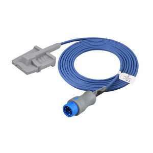
| Soft finger / silicone (reusable) | Adults and pediatrics, longer-term monitoring | Comfortable; more secure fit; better for continuous use | Requires periodic replacement |
| Wrap / Y-type (reusable) | Neonates, infants, small children | Flexible; adapts to small digits; secure wrap | Verify sizing for patient weight range |
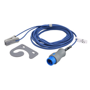
| Ear clip (reusable) | Adults with poor peripheral perfusion or cold extremities | Faster response; less affected by vasoconstriction | Can be uncomfortable for extended periods |
| Adhesive (disposable) | All ages; ICU, surgery, long-term monitoring | Most secure; lowest motion artifact; single-patient infection control | Single use; higher per-patient cost |
For comfortable, secure continuous monitoring on adult and pediatric wards, a reusable soft silicone sensor is the workhorse choice — it stays in place better than a rigid finger clip and is gentler over long wear times, while remaining far more economical than disposables over its service life.
🏭 MedLinket SpO2 sensor compatibility
MedLinket manufactures reusable SpO2 sensors and disposable SpO2 sensors compatible with major monitor brands, including Philips, GE Healthcare, Mindray, Masimo, Nellcor, Nihon Kohden, Comen, Biolight, and Edan. Sensors are manufactured under an ISO 13485:2016 quality system, and the range is FDA 510(k) cleared and CE marked. (Compatibility implies no OEM or endorsement relationship with the monitor brands listed; always confirm the exact match for your monitor model.)
For long-duration and neonatal monitoring, MedLinket offers an over-temperature protection sensor design: the probe contains a temperature sensor that stops the probe and triggers a monitor alarm if skin temperature exceeds 41°C, then automatically resumes SpO2 monitoring once the temperature falls back below 41°C — a safety feature intended to reduce the risk of sensor-related skin injury. (This reduces risk; it does not eliminate it — periodic skin checks and site rotation remain necessary, and the manufacturer notes that wrap measurement is not recommended on the thicker soles of neonates above ~3.5 kg.)
Not sure which sensor fits your monitor? See: How to Identify Which Cables Your Monitor Needs, or contact us for free compatibility verification.
Cleared
Marked
Certified
Countries
Established
For a broader comparison of OEM vs. compatible sensors — including cost, performance, and warranty considerations — read: OEM vs. Compatible Accessories: What to Know →
Clinical tips: getting accurate SpO2 readings every time
Drawing on feedback from clinical engineers and nurses across the hospitals MedLinket supplies, combined with evidence-based best practices, here are the most impactful things you can do to ensure reliable SpO2 data:
6-step routine for reliable SpO2 monitoring
- Inspect the equipment: check the sensor for visible damage, cracked housing, or a frayed cable. Verify it's compatible with your monitor brand and model. Confirm the SpO2 adapter cable is securely connected.
- Select the right sensor type: match it to the patient population (adult finger clip, pediatric soft wrap, neonatal adhesive) and confirm correct size — too large causes alignment errors; too small causes discomfort and artifact.
- Prepare the site: choose a warm, well-perfused digit. Remove nail polish or artificial nails from the target finger. If extremities are cold, warm the hand or switch to an ear-clip sensor.
- Apply correctly: position the sensor so the LED is centered over the nail bed and the photodetector is directly opposite. Snug but not tight enough to impede blood flow.
- Verify the reading: wait a few seconds for it to stabilize. Confirm a strong, regular pleth waveform. Cross-check that PR matches the ECG HR.
- Monitor and rotate: for continuous monitoring, check the sensor site about every 2 hours and rotate to a different digit to prevent pressure injury — especially important in neonates and patients with compromised circulation.

Frequently asked questions
Q: What is a dangerously low SpO2 level?
SpO2 below 90% is generally considered low and warrants prompt medical evaluation; below 85% is a medical emergency. Clinical context matters — a COPD patient with a baseline of 89% is managed differently from a previously healthy adult at the same level. Always assess the patient alongside the number.
Q: Can SpO2 be too high?
For most adults, 100% is fine. But in premature infants, excessively high oxygen can contribute to retinopathy of prematurity — a potentially blinding condition — so neonatal SpO2 targets are carefully managed, often around 90–95% in preterm infants. In COPD, over-oxygenation (targeting SpO2 well above the individualized goal) can suppress respiratory drive in some patients.
Q: Why does my SpO2 fluctuate?
Minor fluctuations of 1–2% are normal and reflect breathing-cycle variation. Larger swings usually indicate sensor placement issues, motion artifact, or genuine respiratory variation (e.g., sleep-apnea desaturation). Check the pleth waveform — if it's stable, the reading is likely reliable.
Q: Is a 92% oxygen level OK?
For healthy adults, 92% is below the normal range (95–100%) and warrants attention. For COPD patients, 88–92% may be the physician-set target. Context is everything — 92% in a previously healthy 30-year-old with pneumonia is clinically significant, while 92% in a stable COPD patient on home oxygen may be expected. Always consult a healthcare provider.
Q: Why does SpO2 alarm more at night?
Several factors contribute: natural sleep-related drops in respiratory drive, positional changes affecting perfusion, undetected sleep apnea, and sensor movement during sleep. With intermittent monitoring, a true sleep desaturation may be missed — by the time a nurse arrives, the patient has woken and SpO2 has normalized. Continuous monitoring with quality SpO2 sensors captures these nocturnal patterns more reliably.
Q: Does dark nail polish really affect SpO2?
Yes, depending on the color. Dark shades — especially blue, green, and black — can absorb light and produce falsely low readings. Clear and lighter shades generally have minimal impact. The simplest fix is to remove polish from the monitored finger, or rotate the sensor 90° so light passes through the sides of the finger instead of the nail bed. Alternatively, switch to an earlobe or toe.
Q: How do I know if it's a real low SpO2 or just a sensor problem?
Check three things: (1) the pleth waveform — a good waveform with a consistent low reading suggests true hypoxemia; a poor waveform suggests a sensor issue; (2) the patient's appearance — cyanosis, dyspnea, and confusion confirm clinical hypoxia; (3) HR vs. PR correlation — if the ECG heart rate and the pulse-oximeter pulse rate don't match, signal quality is poor. For more detail, see our SpO2 sensor troubleshooting guide.
Related SpO2 products & resources
🛒 MedLinket SpO2 products — compatible with major monitor brands
Manufactured under ISO 13485 · FDA 510(k) cleared · CE marked · product-liability insurance up to USD 5M
- Direct-connect SpO2 sensors — reusable finger clip, soft finger, wrap, and ear-clip sensors
- Disposable SpO2 sensors — adhesive and non-adhesive options for adult, pediatric, infant, and neonatal patients
- SpO2 adapter cables — extension and adapter cables for major monitor brands
- Portable pulse oximeters — COX601 / COX801 with PI, PV, and Bluetooth connectivity
Specific product examples:
- Mindray-compatible adult soft SpO2 sensor ($56.00, 20% off)
- Mindray-compatible adult ear-clip SpO2 sensor ($56.00, 20% off)
- Mindray-compatible neonatal wrap SpO2 sensor ($56.00, 20% off)
- Masimo-compatible SpO2 adapter cable
- Nellcor-compatible neonatal wrap SpO2 sensor
- Comen-compatible adult soft SpO2 sensor
- Biolight-compatible adult soft SpO2 sensor
📖 Related articles in this series
- Hospital Monitor Reading & Accessories Guide (pillar page — start here)
- What Is a Normal Heart Rate on a Hospital Monitor?
- What Do ECG Numbers Mean on a Hospital Monitor?
- Understanding NIBP Readings: Systolic, Diastolic, MAP
- What Is EtCO2 and Why Is It Monitored?
- False Alarms on Patient Monitors: Prevention
- How Do SpO2 Sensors Work?
- How to Choose the Right Disposable SpO2 Sensors
- Complete Guide to Pediatric Pulse Oximeters
- Patient Monitor Accessories: Complete Guide by Parameter Type
- Accessory Replacement Schedule: When to Change
- OEM vs. Compatible Accessories: What to Know
- SpO2 Sensor Troubleshooting: Why Your Readings Are Inaccurate
Need compatible SpO2 sensors for your monitor?
Tell us your monitor brand and model. Our engineering team will verify compatibility and recommend the exact SpO2 sensor you need — free of charge. Samples available, with refund on bulk order.
📧 shopify@medlinket.com💬 WhatsApp: +852 6467 3105
Disclaimer: this guide is for educational and clinical-reference purposes. It does not replace clinical training, institutional protocols, or the advice of qualified healthcare professionals. Always follow your facility's policies and the patient's attending physician for clinical decisions. MedLinket (Shenzhen Med-link Electronics Tech Co., Ltd, established 2004) is a manufacturer of patient-monitoring accessories — not a medical diagnostic-device manufacturer. Brand names are referenced for compatibility only and imply no OEM or endorsement relationship.


