By MedLinket Clinical Education Team | Updated: February 2026 | 14 min read Clinically Reviewed
📚 This article is part of our Hospital Monitor Reading & Accessories Guide — a complete 20-article series covering vital sign parameters, alarm troubleshooting, equipment fixes, and accessory selection. Read the full guide for broader context, or continue here for an in-depth look at heart rate monitoring.
⚡ Quick Answer
A normal heart rate on a hospital monitor is 60–100 beats per minute (BPM) for adults at rest. The value appears as "HR" (heart rate, derived from the ECG) or "PR" (pulse rate, derived from the SpO2 sensor), usually displayed in green. A rate below 60 BPM is called bradycardia; above 100 BPM is called tachycardia. Both may be normal or concerning depending on context — always assess the patient, not just the number.
📋 Key Takeaways
-
Normal resting heart rate is 60–100 BPM for adults. Ranges vary significantly by age — newborns (120–160 BPM) and infants (100–160 BPM) are naturally much faster.
-
HR (from ECG) and PR (from SpO2) usually match. A significant discrepancy suggests either a sensor problem or a clinical issue such as a pulse deficit.
-
Heart rate is the first vital sign to change before clinical deterioration — it shifts before blood pressure does. Monitoring trends is as important as checking absolute values.
-
Athletes and patients on beta-blockers may normally run below 60 BPM without any clinical concern.
-
Accurate heart rate readings depend on quality ECG cables, electrodes, and proper skin preparation — poor contact is the #1 cause of false heart rate alarms.
-
Heart rate should always be interpreted alongside other parameters: SpO2, NIBP, EtCO2, and clinical assessment.
📑 Table of Contents
The heart rate number on a bedside monitor is often the first value clinicians glance at when entering a patient's room — and for good reason. As one cardiology intensivist explained in a clinical interview: heart rate is the first parameter that changes before a deterioration cascade starts, and clinicians who focus only on blood pressure are looking at a late indicator that often changes too late for effective intervention.
Whether you're a nurse on a telemetry floor, a respiratory therapist in an ICU, a biomedical engineer maintaining equipment, or a patient's family member trying to understand what the screen is showing, this guide — created by the MedLinket clinical education team drawing on over 20 years of ECG cable and ECG electrode manufacturing and clinical feedback from 2,000+ hospitals worldwide — will give you a thorough understanding of heart rate on a patient monitor.
Understanding Heart Rate Display on a Hospital Monitor
On bedside monitors from major brands like Philips (IntelliVue series), GE Healthcare (CARESCAPE series), and Mindray (BeneVision series), the heart rate display typically includes these components:
| Attribute | Details |
|---|---|
| Label | "HR" (Heart Rate from ECG) and/or "PR" (Pulse Rate from SpO2). You may see both simultaneously. |
| Color | Green (matching the ECG waveform). Specific shade varies by manufacturer. |
| Units | Beats per minute (BPM) |
| Position | Upper-left area, near or within the ECG waveform section |
| Waveform | ECG waveform (PQRST complex). Each sharp upward spike (R-wave) = one heartbeat. |
| Audible Indicator | Beep with each heartbeat. Pitch often correlates with SpO2 level — higher pitch = higher O2 saturation. |
For a complete visual walkthrough of every parameter on a bedside monitor, see our pillar guide: Hospital Monitor Reading & Accessories Guide →
How Heart Rate Is Measured: ECG vs. Pulse Oximetry
A hospital monitor obtains heart rate through two independent pathways. Understanding these helps explain why you might see two different numbers on the same screen — and what it means when they don't agree.
Method 1: ECG-Derived Heart Rate (HR)
The electrocardiogram (ECG) measures the heart's electrical activity through electrodes placed on the patient's skin. These electrodes detect the tiny electrical impulses generated by the cardiac conduction system as it triggers each heartbeat. The signal travels through the conduction pathway: SA node → atria (P wave) → AV node → Bundle of His → bundle branches → Purkinje fibers → ventricles (QRS complex). The monitor counts QRS complexes per minute to produce the HR value.
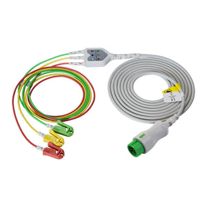
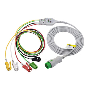
Lead configurations for heart rate monitoring include: 3-lead ECG for basic rhythm monitoring (RA, LA, LL electrodes), 5-lead ECG adding RL ground and one precordial lead for better arrhythmia detection and ST monitoring (standard for ICU/telemetry), and 12-lead ECG for full diagnostic cardiac assessment.
Method 2: SpO2-Derived Pulse Rate (PR)
The pulse oximeter measures the mechanical pulse — the physical expansion of arteries with each heartbeat — as part of its SpO2 measurement. Each time the heart contracts and pushes a bolus of blood through the capillary bed under the SpO2 sensor, the light absorption pattern changes. The monitor counts these pulsations to generate the PR value.
✓ Key Distinction: ECG measures electrical activity (the heart's command to beat), while pulse oximetry measures mechanical activity (the actual pulse wave arriving at the periphery). In a healthy patient with normal cardiac output, these should be virtually identical. When they diverge, it's clinically meaningful — see the HR vs. PR section below.
Normal Heart Rate Ranges by Age Group
Heart rate is profoundly age-dependent. A newborn's heart beats roughly twice as fast as an adult's — and what would be considered tachycardia in an adult is perfectly normal for an infant. The following table provides clinically accepted ranges used in patient monitoring across hospital settings.
| Age Group | Normal Resting HR | Bradycardia (<) | Tachycardia (>) | Clinical Notes |
|---|---|---|---|---|
| Premature Neonate | 120–170 BPM | <120 | >170 | Highly variable; desaturation events common |
| Full-Term Newborn | 120–160 BPM | <120 | >160 | May reach 180+ during crying |
| Infant (1–12 mo) | 100–160 BPM | <100 | >160 | Decreases gradually as child grows |
| Toddler (1–3 yr) | 90–150 BPM | <90 | >150 | Activity and fever significantly raise rate |
| Child (4–10 yr) | 70–120 BPM | <70 | >120 | Approaching adult range |
| Adolescent (11–17 yr) | 60–100 BPM | <60 | >100 | Same as adult ranges |
| Adult (18+ yr) | 60–100 BPM | <60 | >100 | Most commonly referenced range |
| Trained Athlete | 40–60 BPM | — | — | Physiological bradycardia — normal, efficient cardiac function |
| Elderly (>65 yr) | 60–100 BPM | <60 | >100 | Higher arrhythmia risk; medication effects common |
⚠️ Clinical Reminder: For the highest accuracy, assess the heart rate for a full 60 seconds and compare the ECG-derived HR with a manually palpated radial or apical pulse. This is especially important in patients with irregular rhythms (e.g., atrial fibrillation), where monitors may under- or over-count due to variable R-R intervals.
HR vs. PR: What's the Difference and Why It Matters
If you see both "HR" and "PR" on the same monitor screen, they usually show the same number — but when they differ, it's clinically meaningful.
| Feature | HR (Heart Rate) | PR (Pulse Rate) |
|---|---|---|
| Source | ECG (electrical signal) | SpO2 sensor (mechanical pulse) |
| What It Measures | Cardiac electrical depolarizations/min | Arterial pulsations/min at periphery |
| Required Hardware | ECG cables + electrodes | SpO2 sensor + adapter cable |
| Normal State | HR ≈ PR (should match within 1–3 BPM) | |
| Display Color (typical) | Green | Cyan / Light Blue |
💡 Clinical Pearl — The HR-to-PR Cross-Check: Experienced ICU clinicians use the HR-to-PR comparison as a quick reliability check every time they glance at the monitor. If both numbers match, the data is likely accurate. If HR reads 85 but PR reads 62, either the SpO2 sensor has lost adequate perfusion signal (check the pleth waveform and PI), the ECG is double-counting (T-wave overcounting), or there's a true pulse deficit — meaning the heart is generating electrical impulses that aren't producing effective mechanical output. Each scenario requires different action. This cross-check takes seconds and can prevent both missed emergencies and unnecessary interventions.
Bradycardia: When the Heart Rate Is Too Low
Bradycardia is defined as a heart rate below 60 BPM in adults. However, "low" is relative — a sleeping adult or a well-conditioned athlete may have a resting heart rate of 40–55 BPM with no symptoms whatsoever.
When Bradycardia Is Usually Normal
During deep sleep (HR can drop to 40–50 BPM in healthy adults), in trained athletes with high cardiac efficiency, as an expected pharmacological effect of beta-blockers, calcium channel blockers, or digoxin, and in young healthy individuals with high vagal tone.
When Bradycardia Requires Attention
| Cause | Other Signs to Look For | Action |
|---|---|---|
| Heart block (1st, 2nd, 3rd degree) | Prolonged PR interval or dropped QRS on ECG | 12-lead ECG; may require pacing |
| Hypothyroidism | Fatigue, cold intolerance, weight gain | Thyroid function labs |
| Medication overdose | Hypotension, altered consciousness | Review medications; atropine if symptomatic |
| Hyperkalemia | Peaked T waves on ECG, muscle weakness | Stat potassium level; calcium gluconate if critical |
| Cushing reflex (↑ ICP) | Hypertension + bradycardia + irregular breathing | Neurological emergency — immediate escalation |
⚠️ Critical Threshold: A heart rate below 40 BPM with symptoms (hypotension, dizziness, altered consciousness, chest pain) is a medical emergency. Per ACLS guidelines, symptomatic bradycardia may require atropine, transcutaneous pacing, or a temporary pacemaker.
Tachycardia: When the Heart Rate Is Too High
Tachycardia is defined as a heart rate exceeding 100 BPM in adults. Like bradycardia, context is everything — a post-surgical patient with mild tachycardia from pain is very different from a patient in unstable ventricular tachycardia.
| Common Cause | Typical HR Range | Other Signs | Primary Response |
|---|---|---|---|
| Pain | 100–130 BPM | Patient report, guarding | Pain assessment; analgesia |
| Fever / Infection / Sepsis | 100–140 BPM | Elevated temp, WBC changes | Culture, antibiotics, fluids |
| Dehydration / Hypovolemia | 100–130 BPM | Dry mucosa, low urine output | IV fluid resuscitation |
| Anxiety / Stress | 100–120 BPM | Patient appears anxious | Reassurance, anxiolysis |
| Hemorrhage | 110–150+ BPM | Hypotension, pallor, low Hb | Fluid/blood products, surgical consult |
| Atrial Fibrillation (AF) | 100–180 BPM | Irregularly irregular rhythm | Rate control (beta-blocker/CCB) |
| Ventricular Tachycardia (VT) | >150 BPM | Wide QRS (>0.12s), hemodynamic instability | Emergency. Check pulse. Unstable → cardioversion. Pulseless → defibrillation + CPR per ACLS. |
For every 1°C rise in body temperature, heart rate increases by approximately 8–10 BPM. This fever-tachycardia relationship is one of the most predictable vital sign correlations in clinical practice and helps distinguish infection-driven tachycardia from other causes.
⚠️ Priority Threshold: ICU experts recommend continuous ECG monitoring whenever HR exceeds 120 BPM (or 150 BPM in patients without known cardiac disease). A sustained rate above 150 BPM significantly increases myocardial oxygen demand and can precipitate ischemia — especially in patients with coronary artery disease.
What Causes Heart Rate Changes on a Monitor?
Normal / Expected Variations
Respiratory sinus arrhythmia is the most common "normal" variation — HR increases slightly during inspiration and decreases during expiration. This is a normal physiological phenomenon, especially prominent in younger patients. It is not pathological. Other expected variations include sleep-wake cycle changes (HR naturally drops during sleep), positional changes and coughing (transient 10–20 BPM elevation), temperature fluctuations (8–10 BPM increase per 1°C), and medication effects (beta-blockers decrease HR; albuterol, epinephrine, atropine increase it).
Clinically Significant Changes
Four patterns warrant immediate clinical evaluation: (1) an acute HR rise without obvious cause — may be the earliest sign of sepsis, hemorrhage, pulmonary embolism, or acute pain; (2) a new arrhythmia — sudden irregular rhythm, VT, or AF requires immediate ECG interpretation; (3) HR-BP divergence — rising HR with falling blood pressure is an ominous sign of compensatory shock; (4) sudden bradycardia in a non-athletic patient — may indicate vagal stimulation, medication effect, heart block, or raised intracranial pressure.
Why Heart Rate Trends Matter More Than Single Values
A single heart rate reading is a snapshot. What tells the clinical story is the trend. Heart rate is critical in two ways — predicting deterioration and evaluating therapy response. Blood pressure, by contrast, is a measure of cardiac output that changes late in the course of deterioration.
📈 Heart Rate Trend Interpretation Guide
-
Gradually rising HR over hours: Think developing infection (sepsis), worsening pain, dehydration, or progressive bleeding. This is one of the earliest warning signs of patient deterioration.
-
Sudden spike (e.g., 80 → 140 BPM): Consider acute pain, pulmonary embolism, new arrhythmia, hemorrhage, or anxiety/agitation. Requires immediate assessment.
-
HR decreasing after intervention: If HR drops from 130 to 90 after fluid resuscitation, the intervention is working. This is how clinicians use HR trends to evaluate therapy effectiveness.
-
HR not responding to treatment: If HR remains elevated despite fluids and pain management, consider cardiac tamponade, tension pneumothorax, or medication-resistant arrhythmia.
Most modern monitors from Philips, GE, and Mindray offer trend displays that graph vital signs over 1-, 4-, 8-, or 24-hour intervals. Make it a habit to review these trends at the start of each shift and during reassessments.
False Heart Rate Alarms: Causes and Fixes
Heart rate alarms are among the most frequent alarm types in hospitals. Studies have shown that up to 99.4% of cardiac monitor alarms may be false or clinically non-actionable — a major contributor to alarm fatigue, which The Joint Commission has ranked among the top 10 patient safety concerns.
| False Alarm Cause | What Happens | How to Fix |
|---|---|---|
| Loose / Dry ECG Electrode | Loss of contact → "Leads Off" or erratic HR | Replace with fresh ECG electrodes. Change every 24h. |
| Patient Movement / Motion Artifact | Muscle signals mimic cardiac signals → false tachycardia | Secure cables; wait for patient to settle; confirm with manual pulse. |
| T-Wave Overcounting | Monitor mistakes tall T-waves for QRS → displays double the actual rate | Change monitored lead (e.g., Lead II → Lead I). Adjust QRS sensitivity. |
| 50/60 Hz Electrical Interference | Nearby equipment creates noise → false high-frequency activity | Check grounding; move devices away; use shielded ECG cables. |
| Diaphoresis (Sweating) | Moisture reduces electrode adhesion → baseline drift and false alerts | Dry skin, re-prep with alcohol. Use electrodes with strong adhesive. |
| Alarm Limits Too Narrow | Default thresholds trigger on insignificant variations | Customize per patient (e.g., low-limit 50 for athlete on beta-blocker). |
The single most common cause of false heart rate alarms is poor electrode contact. Fresh, low-impedance electrodes with strong adhesive dramatically reduce both "Leads Off" alerts and motion-induced false readings — which is why a 24-hour electrode change schedule is one of the highest-yield, lowest-cost interventions for reducing alarm fatigue on a unit.
For detailed troubleshooting of ECG-related artifacts and leads-off alarms, see: ECG Leads Off Alarm: How to Fix →
How ECG Accessories Affect Heart Rate Accuracy
The accuracy of the heart rate number on your monitor is only as good as the signal chain delivering data from the patient's skin to the processor. That chain consists of three critical components — and weakness in any one degrades the entire reading.
🔗 The ECG Signal Chain — From Patient to Monitor
-
ECG Electrodes (Patient Contact): The electrode converts the body's ionic electrical signal into an electronic signal. Electrode quality directly determines signal-to-noise ratio. MedLinket's electrodes are tested against the YY/T 0196-2005 standard with results that significantly exceed requirements: AC impedance 109Ω (standard: ≤2,000Ω), DC offset voltage 4.11mV (standard: ≤100mV), and internal noise 49.5μV (standard: ≤150μV). Lower values = cleaner signal = more accurate heart rate detection = fewer false alarms.
-
ECG Leadwires (Signal Transmission): Connect individual electrodes to the trunk cable. Damaged, frayed, or corroded leadwires introduce noise, baseline drift, and intermittent signal loss — all of which produce false heart rate readings.
-
ECG Trunk Cable (Connection to Monitor): Connects the leadwire assembly to the monitor. Must match the monitor's specific connector type and pin configuration. A worn trunk cable can cause intermittent disconnections that the monitor interprets as asystole or arrhythmia events.
🏭 MedLinket ECG Products for Reliable Heart Rate Monitoring
All MedLinket ECG cables, ECG leadwires, and ECG electrodes are manufactured in our ISO 13485-certified facility (3 factories: Shenzhen, Shaoguan, Indonesia), 100% factory-tested, and compatible with all major patient monitor brands.
-
Mindray 3-Lead ECG Cable — Pinch/Grabber, IEC ($48.00, 20% off)
-
Mindray 5-Lead ECG Cable — Pinch/Grabber, IEC ($56.00, 20% off)
Compatible brands: Philips · GE Healthcare · Mindray · Nihon Kohden · Dräger · and 30+ more. All products CE marked and FDA 510(k) cleared.
Not sure which ECG cable fits your monitor? See: How to Identify Which Cables Your Monitor Needs, or contact us for free compatibility verification.
FDA 510(k) 19 Clearances
CE 48 Class II
ISO 13485 Certified
120+ Countries
Since 2004 Est.
🛡️ MedLinket Innovation: Eccentric (Off-Center) ECG Electrodes
Standard center-connection electrodes are prone to motion artifact from clothing friction and cable pull. MedLinket's patented eccentric electrode design (Patent CN202120112524.5) places the leadwire connection point off-center with a flexible FPC base and narrow neck structure, allowing 360° cable rotation without disturbing the electrode-skin interface.
Lab-tested results show dramatic improvements: standard center electrodes exhibit baseline drift up to 7,000μV under pull-force simulation, while eccentric electrodes remain virtually unaffected. Under 1N pull-force, center electrodes drop 2,000–7,000μV with incomplete recovery — eccentric electrodes show only a temporary 1,000μV drop with full recovery in 0.1 seconds. These improvements translate directly to fewer false heart rate alarms and better arrhythmia detection accuracy.
For a broader comparison of OEM vs. compatible accessories — including cost analysis and warranty implications — read: OEM vs. Compatible Accessories: What to Know →
Frequently Asked Questions
Q: Why is the heart rate number changing constantly on the monitor?
A: This is normal. Heart rate is a living, dynamic parameter that fluctuates with each breath (respiratory sinus arrhythmia), changes in autonomic tone, and minor positional adjustments. Fluctuations of 3–5 BPM are physiologically normal and expected. The monitor is simply reflecting real-time cardiac activity.
Q: My monitor shows HR 120 but the patient looks fine — should I be worried?
A: Context matters. Check if the patient recently walked, is in pain, has a fever, or is anxious — all can cause transient tachycardia. Also check for artifact: a loose electrode or cable movement can create false high readings. Confirm with a manual pulse check and assess the ECG waveform quality. If the rate is persistent and unexplained, escalate.
Q: Is a resting heart rate of 55 dangerous?
A: Not necessarily. A resting HR of 55 BPM is common in athletes, young healthy adults, and patients taking beta-blockers. It's only concerning if accompanied by symptoms — dizziness, fatigue, syncope, or hypotension. Always assess the patient, not just the number.
Q: Why does my patient's HR and PR show different numbers?
A: If HR (from ECG) and PR (from SpO2) differ by more than 5 BPM, check three things: (1) Is the SpO2 sensor properly placed with a good pleth waveform? (2) Is the ECG double-counting (T-wave overcounting)? (3) Is there a true pulse deficit — the heart is beating but some beats aren't producing effective mechanical output? Each requires different action.
Q: How do I stop false heart rate alarms on my unit?
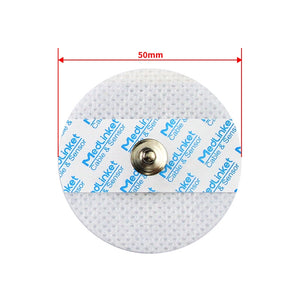
A: Focus on three areas: (1) Electrode quality — use fresh ECG electrodes every 24 hours with proper skin prep (our adult 4.0mm snap electrodes are $11.90/bag of 50, 21% off); (2) Alarm limits — customize thresholds per patient rather than using defaults; (3) Cable quality — replace damaged ECG cables and leadwires on schedule. See our accessory replacement schedule for timing guidelines.
Q: What heart rate requires immediate emergency response?
A: Immediate emergency response for: sustained HR >150 BPM with hemodynamic instability, HR <40 BPM with symptoms, ventricular tachycardia (wide QRS, >150 BPM), pulseless electrical activity (HR on monitor but no pulse), or any rate accompanied by chest pain, altered consciousness, or severe hypotension.
Q: Does the quality of the ECG cable really affect the heart rate reading?
A: Absolutely. The entire ECG signal chain — electrodes, leadwires, trunk cable — directly impacts the quality of the waveform from which heart rate is derived. A degraded cable can introduce noise that causes the monitor to miscount heartbeats or trigger false arrhythmia alarms. This is why hospitals that switch to high-quality compatible ECG accessories often see a measurable reduction in false alarm rates.
Related Articles & Products
🛒 MedLinket ECG Products — Compatible with 30+ Monitor Brands
All products manufactured under ISO 13485 | FDA 510(k) Cleared | CE Marked | $5M Product Liability Insurance
-
One-Piece ECG Cables — Direct-connect 3-lead, 5-lead, and 12-lead
-
ECG Trunk Cables — Connect leadwire sets to monitors
-
ECG Leadwires — Individual lead sets, snap/grabber/pinch options
-
Disposable ECG Electrodes — Standard and eccentric (off-center) designs
-
Holter / ECG Accessories — Holter cables and specialized monitoring
📖 Related Articles in This Series
-
Hospital Monitor Reading & Accessories Guide (Pillar Page — Start Here)
-
Patient Monitor Accessories: Complete Guide by Parameter Type
Need Compatible ECG Cables or Electrodes for Your Monitor?
Tell us your monitor brand and model. Our engineering team will verify compatibility and recommend the exact ECG cable, leadwire, and electrode you need — free of charge. Samples available with refund on bulk order.
📧 shopify@medlinket.com
💬 WhatsApp: +852 6467 3105
Disclaimer: This guide is intended for educational and clinical reference purposes. It does not replace clinical training, institutional protocols, or the advice of qualified healthcare professionals. Always consult your facility's policies and the patient's attending physician for clinical decisions. MedLinket (est. 2004, NEEQ: 833505) is a manufacturer of patient monitoring accessories — not a medical diagnostic device manufacturer.
Declaration: All product names, brands, and OEM part numbers (e.g., Philips, GE, Mindray, and associated model numbers) are used for identification and compatibility-reference purposes only and do not imply any affiliation, partnership, or endorsement. MedLinket products are compatible alternatives independently verified against publicly available specifications. Product images and actual objects may differ slightly in appearance (e.g., connector design, color) but function the same.


