By MedLinket Clinical Education Team | Reviewed by Biomedical Engineering Dept. | Updated: February 2026 | 12 min read
Quick Answer: False alarms occur when patient monitors alert for non-clinical reasons—a loose ECG electrode misread as asystole, an SpO2 sensor artifact flagged as hypoxemia, or a poorly fitting NIBP cuff triggering a blood pressure alarm. Research consistently shows that 85–99% of hospital monitor alarms are false or clinically insignificant. Reduce false alarms through five evidence-based strategies: (1) proper electrode and sensor placement, (2) patient-specific alarm limit customization, (3) daily electrode replacement coordinated with bathing, (4) routine cable and sensor maintenance, and (5) ongoing staff education on patient monitoring systems.
📚 This article is part of the Hospital Monitor Reading & Accessories Guide series.
Related: ECG Leads Off Alarm: How to Fix · Blood Pressure Alarm Troubleshooting · Patient Monitor Accessories Guide
Walk into any ICU, telemetry unit, or PACU and you will hear it—a relentless stream of beeps, chimes, and alerts from cardiac monitors, pulse oximeters, infusion pumps, and ventilators. These alarms are designed to protect patients. But when the vast majority are false, they create a dangerous paradox: the system meant to keep patients safe becomes a threat to their safety.
This is alarm fatigue—and it is not a minor annoyance. The ECRI Institute ranks it among the top 10 health technology hazards, and the Joint Commission has made alarm management a National Patient Safety Goal (NPSG.06.01.01). A 2019 FDA report identified cardiac monitor alarms as the leading cause of alarm-related patient deaths. Each time clinical work is interrupted by a false alarm, the probability of making a clinical error increases by approximately 25%.
This guide explains why false alarms happen across every monitored parameter, how they endanger patients, and what your unit can do to measurably reduce them—drawing on over 20 years of patient monitor accessories manufacturing experience at MedLinket and published clinical evidence.
Table of Contents
- The False Alarm Problem: By the Numbers
- Common Causes of False Alarms by Parameter
- 5 Evidence-Based Strategies to Reduce False Alarms
- How to Tell a Real Alarm from a False Alarm
- The Role of Equipment Quality in Alarm Reduction
- Building a Unit-Level Alarm Reduction Action Plan
- Frequently Asked Questions
The False Alarm Problem: By the Numbers
Before discussing solutions, it is worth understanding the scale of this problem. The data from multiple peer-reviewed studies paints a consistent—and sobering—picture.
| Metric | Finding |
|---|---|
| False/non-actionable alarm rate | 85–99% of ECG monitor alarms are false or clinically insignificant |
| Alarms per bed per day | ~187 audible alarms in a typical ICU |
| Alarms per nurse per shift | ~1,000 across all devices |
| Alarm-related deaths | 566 reported to FDA between 2005–2008 |
| Nursing response rate | 30% of all alarms; only 59% of red (critical) alarms receive response |
↔ Swipe the table sideways to see all columns
A landmark ICU study at the University of California San Francisco documented over 2.5 million unique alarms in just 31 days across 461 patients—including 1.15 million arrhythmia alarms, 612,927 parameter alarms, and 791,632 technical alarms. Of 12,671 annotated arrhythmia alarms, 88.8% were false positives.
The human cost extends beyond staff. Patients in alarm-heavy environments experience sleep deprivation, increased sympathetic nervous system activity, higher re-hospitalization rates, and even increased opioid requirements. Understanding what each alarm means—and which ones are genuine—starts with knowing how to read a hospital monitor accurately.
Common Causes of False Alarms by Parameter
False alarms originate from different sources depending on which monitoring parameter is affected. Understanding the cause by parameter helps you target interventions effectively.
ECG / Heart Rate False Alarms
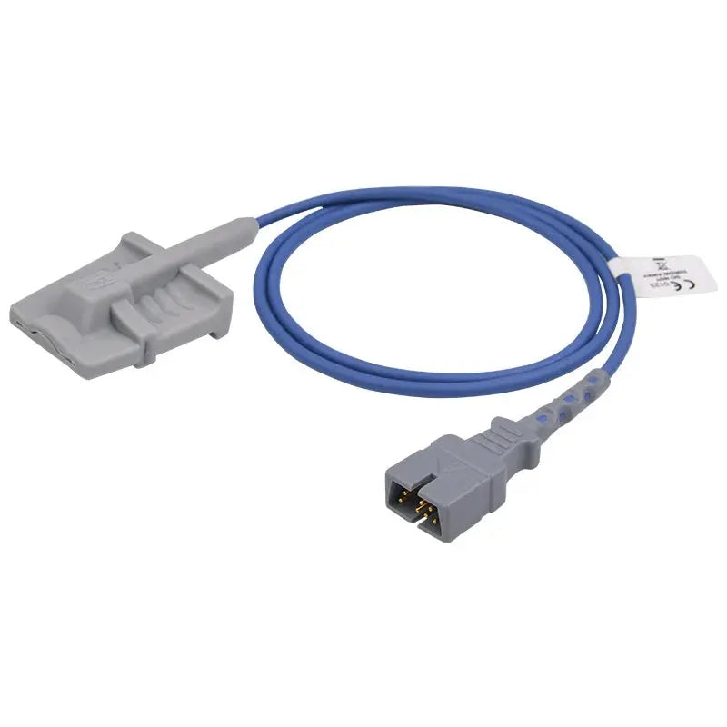
ECG monitoring generates the highest volume of false alarms in most hospital settings. The primary culprits are signal quality issues caused by ECG electrode and cable problems.
| Cause | What the Monitor May Misinterpret | Solution |
|---|---|---|
| Loose or dried-out ECG electrodes | "Leads Off," false asystole, noise mimicking VFib | Replace electrodes every 24h; proper skin prep |
| Patient movement / muscle artifact | False VTach, SVT, or elevated heart rate | Place electrodes over bone (not muscle); secure cable routing |
| 50/60 Hz electrical interference | Baseline noise mimicking atrial fibrillation | Check grounding; move electronic devices away from patient |
| Worn or damaged ECG cables | Intermittent signal loss, baseline drift | Inspect cables regularly; replace ECG lead wires every 6–12 months |
| Inappropriate default alarm limits | Alarm for HR 55 in a fit patient (normal for them) | Customize alarm parameters to the individual patient |
↔ Swipe the table sideways to see all columns
For detailed ECG alarm troubleshooting, see our guides on ECG Leads Off alarm fixes, ECG artifact troubleshooting, and common ECG placement mistakes. Understanding what ECG numbers mean on a hospital monitor also helps clinicians distinguish genuine rhythm changes from artifact.
SpO2 False Alarms
SpO2 sensors are the second most common source of false alarms, particularly low-oxygen alerts that trigger when no actual hypoxemia exists.
| Cause | What Happens | Solution |
|---|---|---|
| Poor sensor placement | Weak pleth waveform → false low SpO2 | Reposition sensor; ensure LED aligns with photodetector |
| Patient movement | Motion artifact → erratic SpO2 values | Consider different site; use motion-tolerant sensor algorithms |
| Cold extremities / poor perfusion | Low perfusion index → unreliable readings | Warm the hand; try ear clip or forehead sensor |
| Nail polish / artificial nails | Light absorption altered → falsely low reading | Remove polish or rotate sensor 90° to side-to-side orientation |
| Ambient light interference | Overhead lights corrupt optical signal | Cover sensor with opaque material |
| Old or damaged SpO2 sensor | Degraded LED/photodetector → chronic unreliability | Replace sensor; inspect SpO2 adapter cables |
↔ Swipe the table sideways to see all columns
For multi-brand compatibility guidance, see compatible SpO2 sensor solutions for multi-brand monitors.
NIBP (Blood Pressure) False Alarms
Blood pressure measurement failures and out-of-range alerts are frequently caused by equipment fit and technique rather than actual hemodynamic instability. For a full troubleshooting walkthrough, see our blood pressure alarm troubleshooting guide.
| Cause | What Happens | Solution |
|---|---|---|
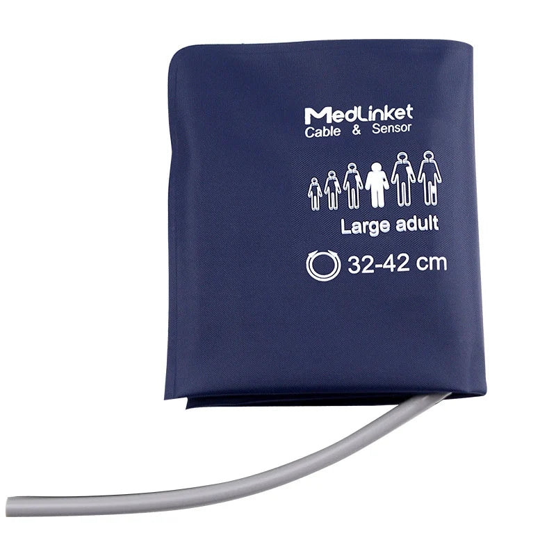
| Wrong NIBP cuff size | Cuff too small → falsely high BP; too large → falsely low BP | Measure arm circumference; select correct cuff per sizing guide |
| Patient movement during measurement | "Measurement failed" alarm or inaccurate reading | Retry when patient is still; educate patient |
| Cuff over clothing | Signal attenuation → failed measurement | Always place cuff on bare skin |
| Kinked or leaking NIBP hose | Incomplete inflation → measurement failure | Inspect hose; replace if damaged |
| Arrhythmias (e.g., atrial fibrillation) | Irregular heartbeat disrupts oscillometric detection | May need manual BP or invasive monitoring |
↔ Swipe the table sideways to see all columns
The single most impactful action for reducing NIBP false alarms is choosing the correct cuff size. A cuff bladder should cover approximately 80% of the patient's arm circumference. For step-by-step technique, see how to put on a blood pressure cuff.
5 Evidence-Based Strategies to Reduce False Alarms
Effective alarm management is not about silencing alarms—it is about eliminating the conditions that generate false ones so that real alarms are noticed and acted upon. These strategies are supported by the AACN, Joint Commission, and AAMI.
Strategy 1: Proper Electrode and Sensor Placement + Skin Preparation
Impact: Reduces ECG false alarms by up to 50% in published quality improvement initiatives.
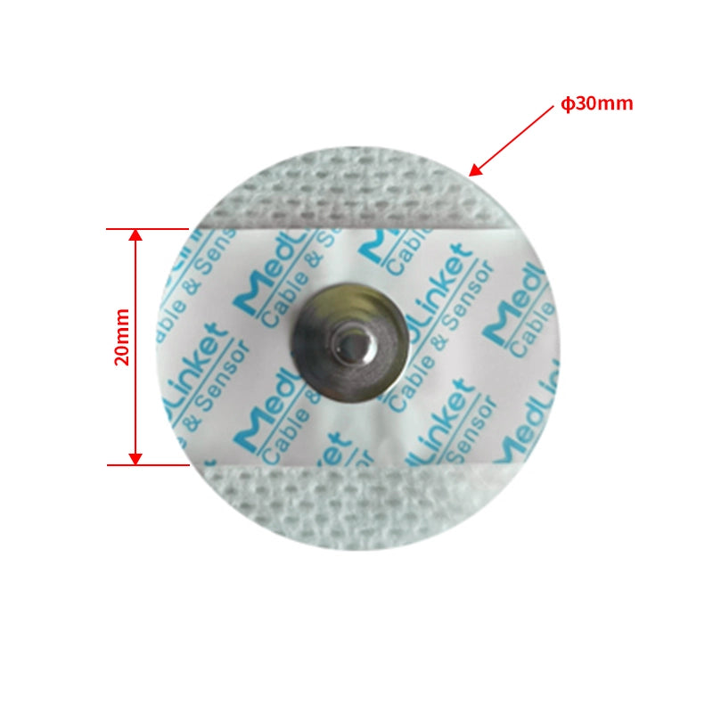
This is the single highest-yield intervention. Waveform artifacts from poor electrode contact are the leading driver of false cardiac alarms. Proper skin preparation—clean with 70% isopropyl alcohol, let dry completely, lightly abrade with gauze, clip (do not shave) chest hair—and correct ECG electrode placement eliminate the signal quality issues that trigger most technical and false arrhythmia alarms.
For SpO2 monitoring, ensure the sensor is properly positioned with the LED and photodetector aligned. For NIBP, use the correct blood pressure cuff size and place the artery marker correctly.
Placement resources: 5-Lead ECG Placement · 3-Lead ECG Placement · ECG Placement Mnemonics · Special Placement Situations
Strategy 2: Customize Alarm Parameters for Each Patient
Impact: One hospital reported a 43% reduction in critical alarms after implementing individualized alarm settings.
Most monitors ship with factory-default alarm limits designed for the general healthy adult population. A resting heart rate of 55 BPM is normal for a young athlete but would trigger a bradycardia alarm on default settings. An SpO2 target of 88–92% is appropriate for many COPD patients but would alarm constantly on a 95% threshold.
Work with your medical team to set patient-specific alarm limits at admission and document them. The AACN's practice alert specifically recommends customizing alarm parameter settings as a core nursing practice.
Strategy 3: Replace Electrodes Daily, Coordinated with Bathing
Impact: Units that standardize bathing and electrode replacement protocols report measurable decreases in false alarms.
Electrode gel degrades over time. After 24 hours, conductive gel dries and adhesion weakens—increasing impedance, causing baseline drift, and triggering "Leads Off" alarms and false arrhythmia detections. Coordinating electrode changes with the patient's daily bath is efficient and effective:
-
Wash the chest with soap and water during bathing
-
Dry thoroughly
-
Apply fresh disposable ECG electrodes to properly prepped skin
-
Reconnect leadwires and verify waveform quality on the monitor
For a complete schedule of when to replace each type of monitoring accessory, see the Accessory Replacement Schedule.
Strategy 4: Routine Cable and Sensor Maintenance
Impact: Eliminates the "invisible" failure mode that causes chronic, hard-to-diagnose alarm problems.
Patient monitor accessories degrade with use—especially under modern high-frequency disinfection protocols. ECG lead wires develop internal micro-fractures from repeated coiling. SpO2 sensor LEDs dim over time. NIBP hoses develop slow leaks. These gradual failures produce chronic, intermittent signal quality issues that generate a steady stream of false alarms.
Establish a preventive maintenance schedule:
| Accessory Type | Inspection Frequency | Replacement Interval |
|---|---|---|
| Disposable ECG electrodes | Every shift | Every 24 hours (every 8h for neonates) |
| ECG leadwires | Every shift (visual) | Every 6–12 months |
| ECG trunk cables | Monthly | Every 2–3 years |
| Reusable SpO2 sensors | Every shift | 12–18 months (per manufacturer) |
| Reusable NIBP cuffs | Weekly | Per condition assessment |
| NIBP hoses | Monthly | Per condition assessment |
| Temperature probes | Before each use | Per condition assessment |
↔ Swipe the table sideways to see all columns
Not sure what cables or sensors your monitor needs? See our guide: How to Identify Which Cables Your Monitor Needs. For cost comparisons between original and compatible replacement accessories, read OEM vs. Compatible Accessories: What to Know.
Strategy 5: Ongoing Staff Education on Monitoring Systems
Impact: Educational interventions that increase clinician competency with monitoring systems measurably decrease alarm frequency.
Training should cover proper skin preparation technique, correct electrode and sensor placement, how to interpret and respond to different alarm types, how to adjust alarm parameters safely, and basic troubleshooting. This is particularly important for new hires and traveling nurses unfamiliar with your unit's specific monitor models.
Training resources: How to Read a Hospital Monitor · How to Read an EKG Quickly · ECG Quality Control System
How to Tell a Real Alarm from a False Alarm
The golden rule of alarm response is: always assess the patient first, then the equipment. But with experience, you can develop a rapid triage instinct. Here is a decision framework:
🔴 Signs It Is a Real Alarm
-
Patient shows clinical signs (cyanosis, distress, altered consciousness, diaphoresis)
-
Waveform is clean and consistent with the alarm
-
Multiple parameters alarming simultaneously (e.g., low SpO2 + tachycardia)
-
Alarm persists despite confirmed good electrode/sensor contact
-
Alarm correlates with recent clinical change (new medication, procedure, deterioration)
⚪ Signs It Is Likely a False Alarm
-
Patient appears comfortable and clinically stable
-
Waveform is noisy, erratic, or flat (artifact or signal loss)
-
Only one parameter alarming while others are normal
-
Alarm coincides with patient movement, position change, or care activity
-
Alarm resolves after repositioning sensor or electrode
⚠️ Never assume an alarm is false without checking the patient. A flat ECG line could be a disconnected electrode or asystole. A "motion artifact" that looks like VTach could be actual VTach in a seizing patient. Always look at the patient before looking at the equipment. If the monitor shows no reading at all, follow the systematic troubleshooting checklist.
The Role of Equipment Quality in Alarm Reduction
While technique and protocols are the foundation, the physical quality of your monitoring accessories directly affects alarm frequency. Low-quality medical cables with poor shielding pick up more electrical interference. Electrodes with inconsistent gel degrade faster. Sensors with subpar optical components produce weaker signals that are more susceptible to motion artifact.
What to Look for in Quality Monitoring Accessories
| Quality Indicator | Why It Reduces False Alarms |
|---|---|
| Braided or foil cable shielding | Blocks 50/60 Hz interference from IV pumps, fluorescent lights, and nearby devices |
| Gold-plated or corrosion-resistant connectors | Maintains low-impedance connection over time, reducing intermittent signal loss |
| Reinforced strain relief at connector points | Prevents internal wire fracture from daily handling, coiling, and cleaning |
| Medical-grade TPU cable jacket | Withstands repeated chemical disinfection without degrading |
| Offset (eccentric) electrode design | Absorbs cable tension away from adhesive area, reducing detachment and baseline drift |
| Consistent gel formulation in electrodes | Maintains low impedance across the full wear period |
↔ Swipe the table sideways to see all columns
💡 Exclusive Data from MedLinket R&D Testing Lab
The following are proprietary test results not available from other sources.
Electrode impedance performance: Per industry standard YY/T 0196-2005, ECG electrodes must maintain AC impedance below 2 kΩ for reliable signal transmission. MedLinket disposable ECG electrodes achieve a tested impedance of just 109 Ω—well below the 2 kΩ threshold—with DC offset of only 4.11 mV (standard requires ≤100 mV) and internal noise of 49.5 μV (standard requires ≤150 μV). Lower impedance means better signal quality, which directly translates to fewer false ECG alarms.
Eccentric electrode motion resistance: In controlled pull-force experiments (1N every 5 seconds), standard concentric ECG electrodes showed baseline drift of 2,000–7,000 μV with incomplete recovery. MedLinket's offset (eccentric) electrodes—protected by Patent CN202120112524.5—showed temporary drift of only ~1,000 μV with full recovery within 0.1 seconds. In click tests (simulated tapping every 4 seconds), concentric electrodes drifted up to 7,000 μV while eccentric electrodes showed no measurable drift. This design is particularly beneficial for Holter ECG monitoring and telemetry applications where patient movement is continuous.
MedLinket also offers a carbon-button electrode variant (V0015H series) that is MRI-safe—producing no induced currents or imaging artifacts—allowing patients to keep electrodes in place during DR, CT, and MRI scans without removal.
Compatible ECG Products by Monitor Brand
| Monitor Brand | 3-Lead ECG Cable | 5-Lead ECG Cable |
|---|---|---|
| Mindray | EA6232A (Grabber) / EA6232B (Snap) | EA6252A (Grabber) / EA6252B (Snap) |
| Philips | M1603A Compatible (Grabber) | Telemetry 5-Lead (Snap) |
| GE Healthcare | 5-Lead Leadwires (Grabber, AHA) | |
| Universal DIN | 10-Lead DIN (Grabber, IEC) | |
↔ Swipe the table sideways to see all columns
Not sure which cable fits your monitor? See How to Identify Which Cables Your Monitor Needs.
About MedLinket — Reducing False Alarms Through Quality Accessories Since 2004
| 🏭 Founded | 2004 · Shenzhen, China (NEEQ: 833505) |
| 🏭 Manufacturing | 3 self-owned factories (Shenzhen, Shaoguan, Indonesia) · 3,500+ molds · 16,651+ product variants |
| 📋 Certifications | ISO 13485 · FDA 510(k) (19 clearances) · CE (48 Class II) · NMPA · MDSAP |
| 🌍 Global Reach | 120+ countries · 2,000+ hospitals including Royal Victoria Hospital (UK) and Institut Hospitalier Jacques Cartier (France) |
| 🔬 Quality | 100% outgoing inspection · $5M product liability insurance · Annual audits by Philips, Mindray, and other OEM partners |
| 🔗 Compatibility | Philips, GE, Mindray, Dräger, Masimo, Nellcor, Nihon Kohden, and 23+ more brands |
Building a Unit-Level Alarm Reduction Action Plan
Sustainable alarm reduction requires a structured approach. The Joint Commission and AACN both recommend an interprofessional alarm management committee. Here is a practical phased framework:
| Phase | Action | Timeline |
|---|---|---|
| 1. Baseline Assessment | Pull alarm data from your monitoring system for 7–14 days. Identify which alarm types are most frequent, peak times, and highest-volume bed locations. | Weeks 1–2 |
| 2. Form Committee | Assemble an interprofessional team: bedside nurses, charge nurse, physician champion, clinical engineer / BMET, and unit manager. | Week 2 |
| 3. Target Top Contributors | Focus on the top 3 alarm types (typically: ECG artifact/leads off, SpO2 low, HR high/low). Implement targeted interventions from the 5 strategies above. | Weeks 3–4 |
| 4. Standardize Protocols | Create unit-standard protocols for electrode replacement timing, skin prep, alarm limit customization at admission, and cable inspection. Integrate into shift handoff. | Weeks 4–6 |
| 5. Educate Staff | Provide hands-on training on proper technique, alarm response triage, and basic equipment troubleshooting. Include in new-hire orientation. | Weeks 5–8 |
| 6. Measure and Iterate | Re-pull alarm data after 30 and 90 days. Compare to baseline. Adjust interventions based on results. | Ongoing |
↔ Swipe the table sideways to see all columns
Pro Tip: When evaluating alarm data, don't just count total alarms—track alarm actionability. The most meaningful metric is what percentage of alarms require clinical intervention versus a technical fix versus no action at all. That ratio reveals whether your alarm system is serving its purpose or generating noise. If equipment-related issues dominate, your accessories may need systematic replacement. If your monitor is having display problems or won't turn on, those hardware issues need to be resolved before alarm optimization can be effective.
Frequently Asked Questions
Q: What percentage of hospital monitor alarms are actually false?
Research consistently shows that 85–99% of ECG monitor alarms are false or clinically insignificant. The exact percentage varies by unit type, patient population, and monitoring setup. Across all monitoring devices (not just ECG), an estimated 72–99% of all alarms do not require a clinical intervention.
Q: How can I tell if an alarm is false or real?
Always assess the patient first—not the monitor. True alarms typically correlate with clinical signs (patient in distress, multiple parameters abnormal, clean waveform showing the alarmed condition). False alarms typically involve noisy or absent waveforms, occur during patient movement or care activities, and resolve when the sensor or electrode is repositioned. See the detailed decision framework above.
Q: Is it safe to silence or disable alarms?
Silencing an alarm temporarily pauses the sound for a set period (typically 1–3 minutes) and is acceptable when you have assessed the patient and are actively fixing the issue. Disabling an alarm turns it off entirely and should be done only with a physician order and clear clinical justification, documented in the patient record. The FDA and Joint Commission have linked alarm disabling to patient deaths. The goal is never to turn off alarms—it is to reduce false alarms so real ones stand out.
Q: Does better equipment really reduce false alarms?
Yes. Quality ECG cables with proper shielding reduce electrical interference. Electrodes with consistent, well-formulated gel maintain lower impedance throughout the wear period. Offset electrode designs reduce motion-induced detachment. These factors complement proper technique—a quality cable will not fix bad skin prep, but it will ensure a well-prepped site delivers a clean signal to the monitor. For OEM vs. compatible accessory comparisons, see our dedicated guide.
Q: What is the Joint Commission's requirement for alarm management?
The Joint Commission established alarm management as a National Patient Safety Goal (NPSG.06.01.01). Accredited hospitals must establish alarm system safety as a priority, identify the most important alarm signals to manage, establish policies and procedures for managing alarms, and educate staff about alarm system management. This has been a compulsory requirement since 2016.
Ready to Reduce False Alarms on Your Unit?
MedLinket offers a full range of ECG cables, disposable ECG electrodes, SpO2 sensors, NIBP cuffs, and temperature probes—all designed for reliable signal quality and compatible with 30+ monitor brands. FDA cleared. CE marked. ISO 13485 certified.
Browse ECG Products →Browse SpO2 Products →
Free compatibility verification & sample requests: shopify@medlinket.com · WhatsApp: +852 6467 3105
📚 Continue Reading: Hospital Monitor Reading & Accessories Guide
A. Parameter Basics:
What is SpO2? · Normal Heart Rate · ECG Numbers Explained · NIBP Readings · What is EtCO2?
B. Alarm Troubleshooting:
ECG Leads Off Alarm · Blood Pressure Alarm · False Alarm Prevention (You are here)
C. Equipment Troubleshooting:
Monitor Shows No Reading · Display Problems · Monitor Not Turning On · When to Call Biomed
D. Accessories:
Accessories by Parameter · Identify Your Cables · OEM vs. Compatible · Replacement Schedule


